File:Keith1921 fig021.jpg: Difference between revisions
From Embryology
No edit summary |
mNo edit summary |
||
| Line 1: | Line 1: | ||
==Fig. 21. Schematic Transverse Sections of two Human Embryos== | |||
A, In the 3rd. week of development. | |||
B, In the 4th week of development. | |||
The numbers are placed on corresponding points : Epiblast, shaded ; hypoblast, black ; mesoblast, stippled. | |||
1. Neural groove and canal. | |||
2. Epiblast of embryo. | |||
3. Epiblast lining amnion. Only the attachment of the amnion is represented in B. | |||
4. Paraxial mesoblast. | |||
5. Intermediate cell mass. | |||
6. Coelom, bounded by the somatopleure externally and splanchnopleure internally. | |||
8. Mesoblast on amnion. | |||
9, 10. Chorion. | |||
11. Notochord. | |||
12. Archenteron. | |||
Revision as of 09:58, 22 December 2014
Fig. 21. Schematic Transverse Sections of two Human Embryos
A, In the 3rd. week of development.
B, In the 4th week of development.
The numbers are placed on corresponding points : Epiblast, shaded ; hypoblast, black ; mesoblast, stippled.
1. Neural groove and canal.
2. Epiblast of embryo.
3. Epiblast lining amnion. Only the attachment of the amnion is represented in B.
4. Paraxial mesoblast.
5. Intermediate cell mass.
6. Coelom, bounded by the somatopleure externally and splanchnopleure internally.
8. Mesoblast on amnion.
9, 10. Chorion.
11. Notochord.
12. Archenteron.
File history
Click on a date/time to view the file as it appeared at that time.
| Date/Time | Thumbnail | Dimensions | User | Comment | |
|---|---|---|---|---|---|
| current | 10:08, 22 December 2014 | 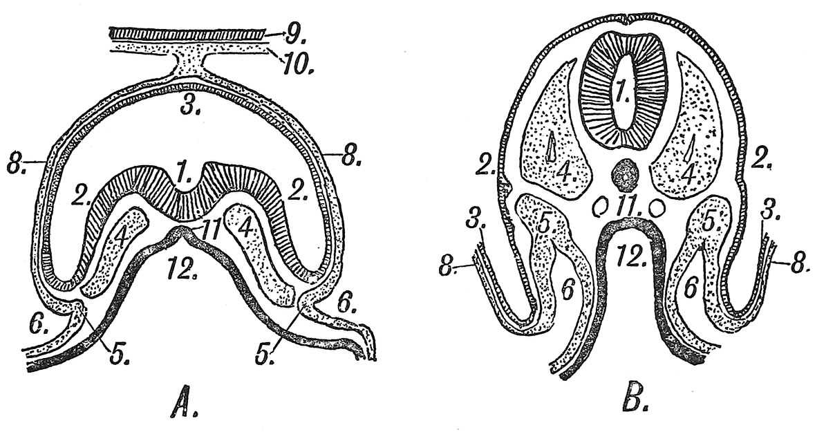 | 1,179 × 625 (160 KB) | Z8600021 (talk | contribs) | |
| 09:48, 22 December 2014 | 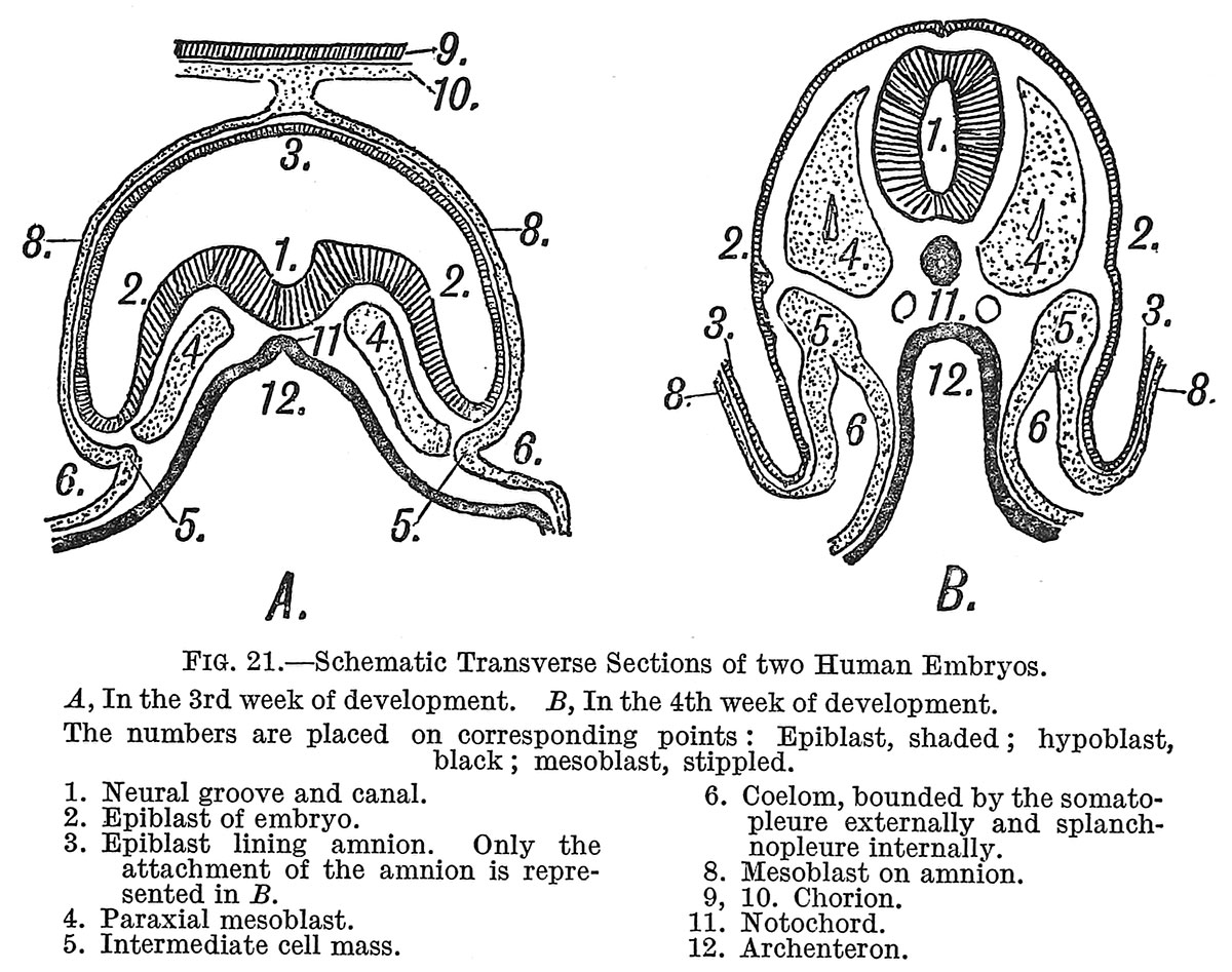 | 1,200 × 947 (254 KB) | Z8600021 (talk | contribs) |
You cannot overwrite this file.
File usage
The following 2 pages use this file: