File:Keith1902 fig078.jpg
Keith1902_fig078.jpg (788 × 600 pixels, file size: 81 KB, MIME type: image/jpeg)
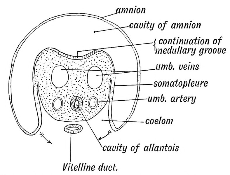
Fig. 78. Diagrammatic section showing the structures which go to form the Umbilical Cord
(After Wilhelm His (1831-1904))
Transverse Section of the Umbilical Cord a section of the cord shows :
- Two umbilical arteries (continuations of the primitive dorsal aortae).
- One umbilical vein, formed by the fusion of the two original veins.
- The cavity of the allantois formed from the hind gut. Within the cord its lumen becomes obliterated early.
- The vitelline duct, the stalk of the yolk sac, communicating with the intestine and yolk sac. It becomes obliterated in the 3rd month.
- Wharton's jelly, a primitive embryonic tissue composed of branching cells in a mucoid matrix.
- A covering of epiblast. The amnion is attached round the placental insertion of the cord.
Up to the end of the 4th week the embryo is closely united to the chorion by the short body-stalk (Fig. 75), but in the second month the cord elongates, and in the third month it measures about 12 cm. and about 40 cm. (16 inches) at birth.
| Historic Disclaimer - information about historic embryology pages |
|---|
| Pages where the terms "Historic" (textbooks, papers, people, recommendations) appear on this site, and sections within pages where this disclaimer appears, indicate that the content and scientific understanding are specific to the time of publication. This means that while some scientific descriptions are still accurate, the terminology and interpretation of the developmental mechanisms reflect the understanding at the time of original publication and those of the preceding periods, these terms, interpretations and recommendations may not reflect our current scientific understanding. (More? Embryology History | Historic Embryology Papers) |
- Foetus and Uterus Connection: Fig. 74. Blastodermic Vesicle Somatopleure | Fig. 75. Somatopleuric Head Fold human ovum of 15 days | Fig. 76. Amnion, Chorion, and Decidua 3rd month | Fig. 77. Placenta formation Elements | Fig. 78. Umbilical Cord Structures | Modern Notes
| Historic Disclaimer - information about historic embryology pages |
|---|
| Pages where the terms "Historic" (textbooks, papers, people, recommendations) appear on this site, and sections within pages where this disclaimer appears, indicate that the content and scientific understanding are specific to the time of publication. This means that while some scientific descriptions are still accurate, the terminology and interpretation of the developmental mechanisms reflect the understanding at the time of original publication and those of the preceding periods, these terms, interpretations and recommendations may not reflect our current scientific understanding. (More? Embryology History | Historic Embryology Papers) |
Human Embryology and Morphology (1902): Development or the Face | The Nasal Cavities and Olfactory Structures | Development of the Pharynx and Neck | Development of the Organ of Hearing | Development and Morphology of the Teeth | The Skin and its Appendages | The Development of the Ovum of the Foetus from the Ovum of the Mother | The Manner in which a Connection is Established between the Foetus and Uterus | The Uro-genital System | Formation of the Pubo-femoral Region, Pelvic Floor and Fascia | The Spinal Column and Back | The Segmentation of the Body | The Cranium | Development of the Structures concerned in the Sense of Sight | The Brain and Spinal Cord | Development of the Circulatory System | The Respiratory System | The Organs of Digestion | The Body Wall, Ribs, and Sternum | The Limbs | Figures | Embryology History
Reference
Keith A. Human Embryology and Morphology. (1902) London: Edward Arnold.
Cite this page: Hill, M.A. (2024, April 27) Embryology Keith1902 fig078.jpg. Retrieved from https://embryology.med.unsw.edu.au/embryology/index.php/File:Keith1902_fig078.jpg
- © Dr Mark Hill 2024, UNSW Embryology ISBN: 978 0 7334 2609 4 - UNSW CRICOS Provider Code No. 00098G
File history
Click on a date/time to view the file as it appeared at that time.
| Date/Time | Thumbnail | Dimensions | User | Comment | |
|---|---|---|---|---|---|
| current | 10:15, 7 January 2014 |  | 788 × 600 (81 KB) | Z8600021 (talk | contribs) |
You cannot overwrite this file.
File usage
The following 2 pages use this file:

