File:Keith1902 fig056.jpg
Keith1902_fig056.jpg (800 × 278 pixels, file size: 49 KB, MIME type: image/jpeg)
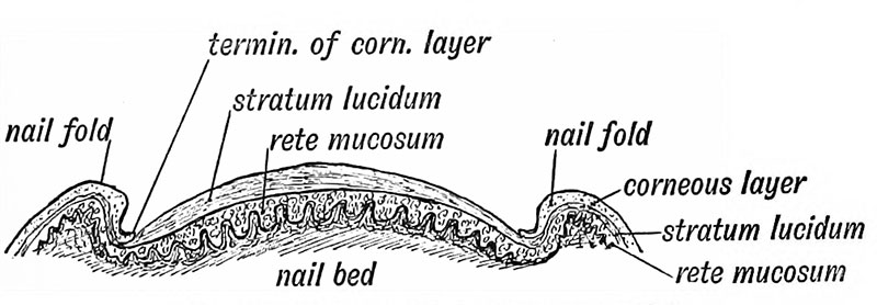
Fig. 56 Diagrammatic Section across a Nail
The nails are made up of the basal, stratum mucosum, and stratum lucidum layers of the skin (Fig. 56), the corneous layer being lost after the 4th month of foetal life.
They appear first in the 3rd month as fields of thickened epidermis on the tips of the digits, but are afterwards shifted dorsally, carrying their palmar nerves with them, so that the terminal phalanx is wholly supplied from the palmar digital branches. The nail of the little toe, a digit in a retrograde phase of development, is frequently shaped like a claw, probably a reversion to a primitive form. The nail is produced on the scattered papillae (the matrix) at its root. The area of production is marked by the lunule. On the nail bed, in front of the lunule, the papillae are arranged in longitudinal rows. If the nail be pressed, as by the boot, the lateral papillae, under the nail fold (see Fig. 56) are directed downwards, and their epithelial outgrowths follow the same direction, thus causing ingrowing nail.
About the end of the 7th month the matrix of the nail root becomes differentiated, active growth sets in and the terminal margin of the nails become free ; it grows forwards over the corneous layer which covers the terminal row of papillae of the nail bed. The ridge of corneous epithelium under the nail-tip represents the hoof of ungulates.
| Historic Disclaimer - information about historic embryology pages |
|---|
| Pages where the terms "Historic" (textbooks, papers, people, recommendations) appear on this site, and sections within pages where this disclaimer appears, indicate that the content and scientific understanding are specific to the time of publication. This means that while some scientific descriptions are still accurate, the terminology and interpretation of the developmental mechanisms reflect the understanding at the time of original publication and those of the preceding periods, these terms, interpretations and recommendations may not reflect our current scientific understanding. (More? Embryology History | Historic Embryology Papers) |
- The Skin and its Appendages: Fig. 51. Skin strata first month | Fig. 52. Skin strata second month | Fig. 53. Skin strata sixth month onwards | Fig. 54. Common dermal papillae patterns on the finger tips | Fig. 55. Developing Hair | Fig. 56. Diagrammatic Section across a Nail | Fig. 57. Stages in Mamma development | Fig. 58. Section of the Breast | Figures
| Historic Disclaimer - information about historic embryology pages |
|---|
| Pages where the terms "Historic" (textbooks, papers, people, recommendations) appear on this site, and sections within pages where this disclaimer appears, indicate that the content and scientific understanding are specific to the time of publication. This means that while some scientific descriptions are still accurate, the terminology and interpretation of the developmental mechanisms reflect the understanding at the time of original publication and those of the preceding periods, these terms, interpretations and recommendations may not reflect our current scientific understanding. (More? Embryology History | Historic Embryology Papers) |
Human Embryology and Morphology (1902): Development or the Face | The Nasal Cavities and Olfactory Structures | Development of the Pharynx and Neck | Development of the Organ of Hearing | Development and Morphology of the Teeth | The Skin and its Appendages | The Development of the Ovum of the Foetus from the Ovum of the Mother | The Manner in which a Connection is Established between the Foetus and Uterus | The Uro-genital System | Formation of the Pubo-femoral Region, Pelvic Floor and Fascia | The Spinal Column and Back | The Segmentation of the Body | The Cranium | Development of the Structures concerned in the Sense of Sight | The Brain and Spinal Cord | Development of the Circulatory System | The Respiratory System | The Organs of Digestion | The Body Wall, Ribs, and Sternum | The Limbs | Figures | Embryology History
Reference
Keith A. Human Embryology and Morphology. (1902) London: Edward Arnold.
Cite this page: Hill, M.A. (2024, April 27) Embryology Keith1902 fig056.jpg. Retrieved from https://embryology.med.unsw.edu.au/embryology/index.php/File:Keith1902_fig056.jpg
- © Dr Mark Hill 2024, UNSW Embryology ISBN: 978 0 7334 2609 4 - UNSW CRICOS Provider Code No. 00098G
File history
Click on a date/time to view the file as it appeared at that time.
| Date/Time | Thumbnail | Dimensions | User | Comment | |
|---|---|---|---|---|---|
| current | 09:14, 7 January 2014 | 800 × 278 (49 KB) | Z8600021 (talk | contribs) |
You cannot overwrite this file.
File usage
The following 3 pages use this file:

