File:Keibel Mall 2 600.jpg
From Embryology
Size of this preview: 800 × 554 pixels. Other resolution: 1,000 × 692 pixels.
Original file (1,000 × 692 pixels, file size: 77 KB, MIME type: image/jpeg)
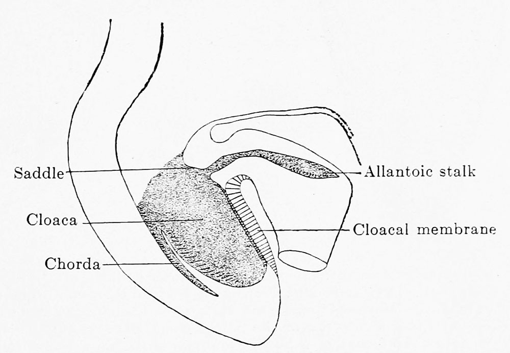
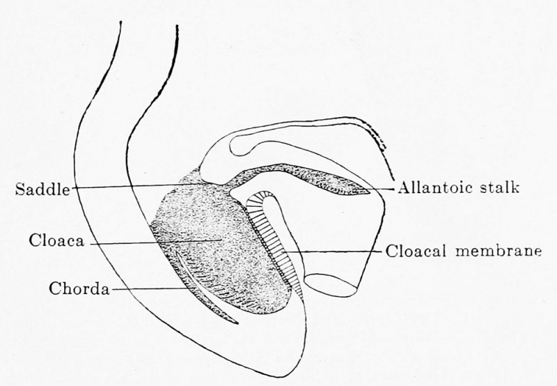
Fig. 600. — Reconstruction of the caudal end of the body of Embryo Template:Pfannenstiel III, 2.6 mm greatest length and with 13-14 pairs of primitive segments. X 50. Cloaca and cloacal membrane have grown greatly in comparison with the conditions in the embryo shown in Fig. 599; the allantoic stalk is given off at a right angle from the cloaca, immediately above the cloacal membrane. The ventral wall of the cloaca is formed only by the cloacal membrane.
File history
Click on a date/time to view the file as it appeared at that time.
| Date/Time | Thumbnail | Dimensions | User | Comment | |
|---|---|---|---|---|---|
| current | 13:35, 12 November 2018 | | 1,000 × 692 (77 KB) | Z8600021 (talk | contribs) | Fig. 600. Reconstruction of the caudal end of the body of Embryo Template:Pfannenstiel III, 2.6 mm greatest length and with 13-14 pairs of primitive segments. X 50. Cloaca and cloacal membrane have grown greatly in comparison with the conditions in the... |
| 13:29, 12 November 2018 | 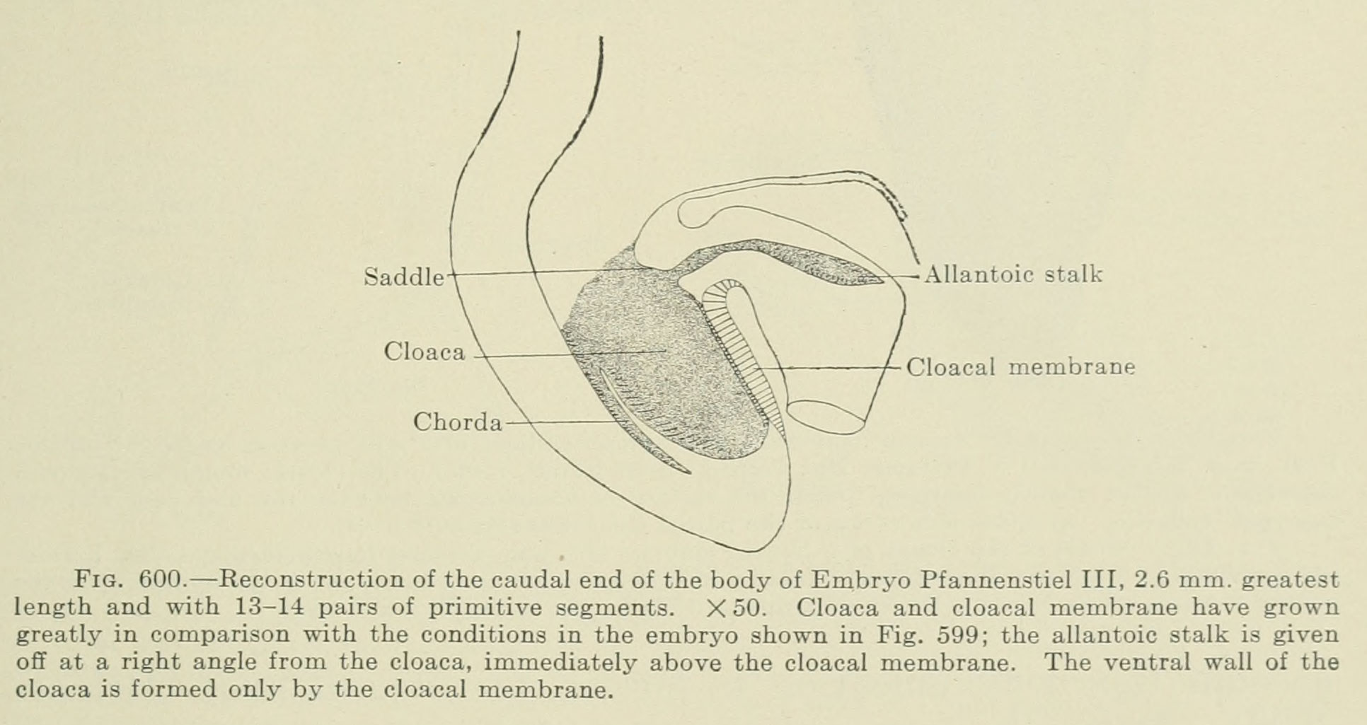 | 1,932 × 1,025 (184 KB) | Z8600021 (talk | contribs) | Fig. 600. — Reconstruction of the caudal end of the body of Embryo Template:Pfannenstiel III, 2.6 mm greatest length and with 13-14 pairs of primitive segments. X 50. Cloaca and cloacal membrane have grown greatly in comparison with the conditions in... |
You cannot overwrite this file.
File usage
The following 3 pages use this file: