File:Keibel Mall 2 306.jpg
From Embryology

Size of this preview: 410 × 600 pixels. Other resolution: 569 × 832 pixels.
Original file (569 × 832 pixels, file size: 66 KB, MIME type: image/jpeg)
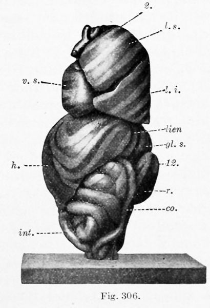
Fig. 306. Lateral view of a model of the viscera from a 65 mm embryo
X 2 diam.
(After C. M. Jackson.)
2, 8, q, 12, ribs; at. d., <it. 8., right and left atria: co., colon; nL s., left suprarenal gland; h., liver; int., small intestine; lien, spleen; /. £., /. m., I. 8., inferior, middle, and superior lobes of the lung; p. v., vermiform process and cecum; r., kidney; v. 8., left ventricle; v. u., umbilical vein.
File history
Click on a date/time to view the file as it appeared at that time.
| Date/Time | Thumbnail | Dimensions | User | Comment | |
|---|---|---|---|---|---|
| current | 15:47, 4 February 2019 |  | 569 × 832 (66 KB) | Z8600021 (talk | contribs) |
You cannot overwrite this file.
File usage
The following 2 pages use this file: