File:Keibel Mall 2 264.jpg
From Embryology
Size of this preview: 279 × 600 pixels. Other resolution: 698 × 1,500 pixels.
Original file (698 × 1,500 pixels, file size: 276 KB, MIME type: image/jpeg)
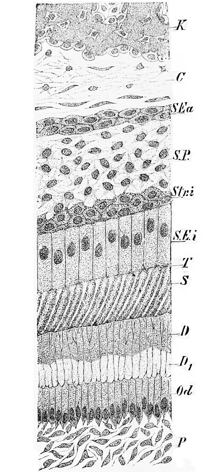
Fig. 264. Section through a developing molar tooth of Didelphys
(After Rose.)
C, connective tissue; D, calcified, and D\, uncalcified dentine; K, wall of dental alveolus; Od, odontoblasts; P, pulp 'ells; S, enamel; SEa, outer epithelial layer of the enamelorgan; S.E.i, ameloblasts; S.P., enamel-pulp; Str.i, intermediate layer of enamel-organ; T, Tomes's processes of the ameloblasts.
File history
Click on a date/time to view the file as it appeared at that time.
| Date/Time | Thumbnail | Dimensions | User | Comment | |
|---|---|---|---|---|---|
| current | 21:29, 2 April 2014 | | 698 × 1,500 (276 KB) | Z8600021 (talk | contribs) | ==Fig. 264. Section through a developing molar tooth of Didelphys== (After Rose.) C, connective tissue; D, calcified, and D\, uncalcified dentine; K, wall of dental alveolus; Od, odontoblasts; P, pulp 'ells; S, enamel; SEa, outer epithelial layer of ... |
You cannot overwrite this file.
File usage
The following 3 pages use this file: