File:Keibel Mall 2 005.jpg
From Embryology

Size of this preview: 655 × 600 pixels. Other resolution: 1,000 × 916 pixels.
Original file (1,000 × 916 pixels, file size: 330 KB, MIME type: image/jpeg)
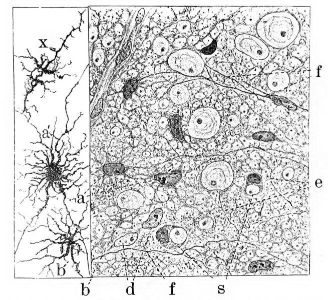
Fig. 5. Combined drawings, after Golgi and Benda methods spinal cord fetal pig 20 cm long
Showing syncytial character of neuroglia framework and the first appearance of neuroglia fibres. a, neuroglia cells after the Benda method; a', similar cell after the Golgi method; e and f, neuroglia fibres beginning to take the neuroglia stain; b, pseudocell due to staining of a portion of the syncytium such as seen at b; s, seal-ring cells.
(After Hardesty.)
File history
Click on a date/time to view the file as it appeared at that time.
| Date/Time | Thumbnail | Dimensions | User | Comment | |
|---|---|---|---|---|---|
| current | 09:35, 3 March 2017 |  | 1,000 × 916 (330 KB) | Z8600021 (talk | contribs) | |
| 09:34, 3 March 2017 |  | 1,415 × 1,219 (434 KB) | Z8600021 (talk | contribs) |
You cannot overwrite this file.
File usage
The following 2 pages use this file: