File:Keibel Mall 2 004.jpg: Difference between revisions
From Embryology
mNo edit summary |
|||
| Line 1: | Line 1: | ||
==Fig. 4. Development of neuroglia framework== | ==Fig. 4. Development of neuroglia framework== | ||
Combined drawing, after Golgi and Benda methods, showing section of spinal cord of embryo pig 30 mm | Combined drawing, after Golgi and Benda methods, showing section of spinal cord of embryo pig 30 mm long. It can be seen that the wealth of communicating processes of the neuroglia cells is not shown in the Golgi portion. | ||
ep, ependymal layer; m, marginal layer; n mantle or nuclear layer; p pia mater. | ep, ependymal layer; m, marginal layer; n mantle or nuclear layer; p pia mater. | ||
(After Hardesty.) | (After Hardesty.) | ||
{{Ref-Hardesty1904a}} | |||
{{Ref-Hardesty1904b}} | |||
Latest revision as of 09:53, 3 March 2017
Fig. 4. Development of neuroglia framework
Combined drawing, after Golgi and Benda methods, showing section of spinal cord of embryo pig 30 mm long. It can be seen that the wealth of communicating processes of the neuroglia cells is not shown in the Golgi portion.
ep, ependymal layer; m, marginal layer; n mantle or nuclear layer; p pia mater.
(After Hardesty.)
Hardesty I. On the development and nature of the neuroglia. (1904) Amer. J Anat. 3.
Hardesty I. On the occurrence of sheath cells and the nature of the axone sheaths in the central nervous system. (1904) Amer. J Anat. 4.
File history
Click on a date/time to view the file as it appeared at that time.
| Date/Time | Thumbnail | Dimensions | User | Comment | |
|---|---|---|---|---|---|
| current | 09:29, 3 March 2017 |  | 1,000 × 722 (199 KB) | Z8600021 (talk | contribs) | |
| 09:29, 3 March 2017 | 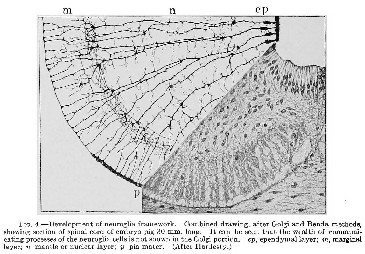 | 1,408 × 979 (288 KB) | Z8600021 (talk | contribs) |
You cannot overwrite this file.
File usage
The following 2 pages use this file: