File:Johnston1907 fig021.jpg
From Embryology
Size of this preview: 709 × 600 pixels. Other resolution: 1,000 × 846 pixels.
Original file (1,000 × 846 pixels, file size: 98 KB, MIME type: image/jpeg)
Summary
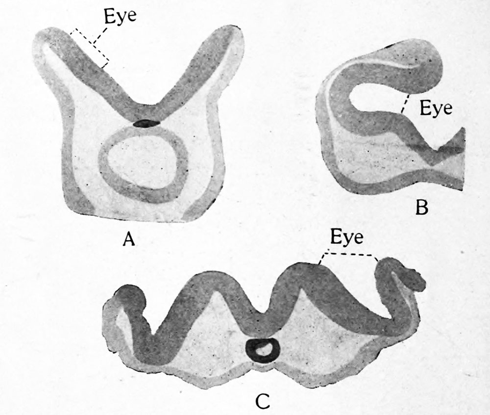
Fig. 21. Transverse sections through the region of the optic vesicles in selachian, avian and mammalian embryos : A, Torpedo ocellata ; B, Callus domesticus ; C, Cavia cobaya. After Froriep. These and the following figure show that the optic vesicles arise from the borders of the neural plate.
File history
Click on a date/time to view the file as it appeared at that time.
| Date/Time | Thumbnail | Dimensions | User | Comment | |
|---|---|---|---|---|---|
| current | 23:06, 23 February 2020 | | 1,000 × 846 (98 KB) | Z8600021 (talk | contribs) | |
| 23:04, 23 February 2020 |  | 2,088 × 1,709 (356 KB) | Z8600021 (talk | contribs) | Fig. 21. Transverse sections through the region of the optic vesicles in selachian, avian and mammalian embryos : A, Torpedo ocellata ; B, Callus domesticus ; C, Cavia cobaya. After Froriep. These and the following figure show that the optic vesicles arise from the borders of the neural plate. |
You cannot overwrite this file.
File usage
The following 2 pages use this file: