File:Johnston1907 fig021.jpg: Difference between revisions
From Embryology
mNo edit summary |
(Z8600021 uploaded a new version of File:Johnston1907 fig021.jpg) |
(No difference)
| |
Latest revision as of 23:06, 23 February 2020
Fig. 21. Transverse sections through the region of the optic vesicles in selachian, avian and mammalian embryos
A, Torpedo ocellata ; B, Callus domesticus ; C, Cavia cobaya.
After Froriep. These and the following figure show that the optic vesicles arise from the borders of the neural plate.
Johnston JB. The Nervous System of Vertebrates. (1907) Blakiston's Son & Co., London.
| Historic Disclaimer - information about historic embryology pages |
|---|
 |
Cite this page: Hill, M.A. (2024, May 13) Embryology Johnston1907 fig021.jpg. Retrieved from https://embryology.med.unsw.edu.au/embryology/index.php/File:Johnston1907_fig021.jpg
- © Dr Mark Hill 2024, UNSW Embryology ISBN: 978 0 7334 2609 4 - UNSW CRICOS Provider Code No. 00098G
File history
Click on a date/time to view the file as it appeared at that time.
| Date/Time | Thumbnail | Dimensions | User | Comment | |
|---|---|---|---|---|---|
| current | 23:06, 23 February 2020 | 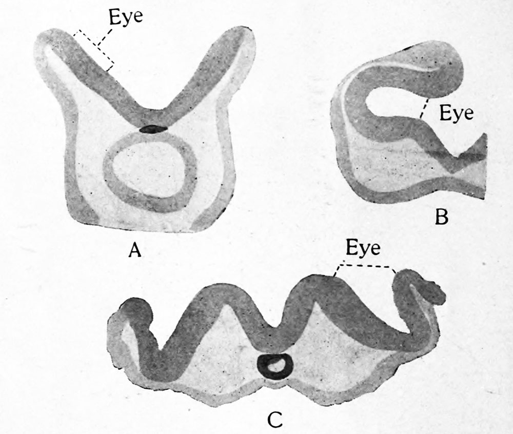 | 1,000 × 846 (98 KB) | Z8600021 (talk | contribs) | |
| 23:04, 23 February 2020 |  | 2,088 × 1,709 (356 KB) | Z8600021 (talk | contribs) | Fig. 21. Transverse sections through the region of the optic vesicles in selachian, avian and mammalian embryos : A, Torpedo ocellata ; B, Callus domesticus ; C, Cavia cobaya. After Froriep. These and the following figure show that the optic vesicles arise from the borders of the neural plate. |
You cannot overwrite this file.
File usage
The following 2 pages use this file: