File:Johnson1914 plate07.jpg

Original file (1,200 × 1,756 pixels, file size: 207 KB, MIME type: image/jpeg)
Plate 7
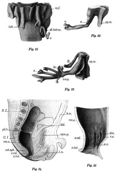
Fig. 21 Lower portion of the primary fold of a human embryo of 135 mm. X 72. gl.intrm., intramuscular gland; sc.f., secondary fold; tub., tubules; x, marks the point where the gland penetrates the internal sphincter muscle.
Fig. 22 Intramuscular gland. Human embryo of 245 mm. X 72. a., acinouslike ending; ep.w., epithelial wall.
Fig. 23 Branched intramuscular gland. Human embryo of 245 mm. X 72. a., acinous-like termination; amp., ampulla of gland; br., branch; ep.w., epithelial wall.
Fig. 24 Sagittal section through the pelvis at birth. X 1. b.w., body wall; bid., bladder; C.I., 1st coccygeal vertebra; ext.sph., external sphincter muscle; La., levator ani muscle; pl.tr., plica transversalis recti; rec.a., rectal ampulla; S.I., 1st sacral vertebra; sym.p., symphysis pubis; z.col., zona columnaris; z.int., zona intermedia.
Fig. 25 Sagittal section through the lower part of the rectum. X 6. a.v., anal valve; r.c, rectal column; rec.a., rectal ampulla; sin., rectal sinus; z.col., zona columnaris; z.int., zona intermedia.
Reference
Johnson FP. The development of the rectum in the human embryo. (1914) Amer. J Anat. 16(1): 1-58.
Cite this page: Hill, M.A. (2024, April 28) Embryology Johnson1914 plate07.jpg. Retrieved from https://embryology.med.unsw.edu.au/embryology/index.php/File:Johnson1914_plate07.jpg
- © Dr Mark Hill 2024, UNSW Embryology ISBN: 978 0 7334 2609 4 - UNSW CRICOS Provider Code No. 00098G
File history
Click on a date/time to view the file as it appeared at that time.
| Date/Time | Thumbnail | Dimensions | User | Comment | |
|---|---|---|---|---|---|
| current | 11:19, 18 November 2016 |  | 1,200 × 1,756 (207 KB) | Z8600021 (talk | contribs) | |
| 11:18, 18 November 2016 |  | 1,487 × 2,270 (272 KB) | Z8600021 (talk | contribs) | ===Plate 7=== Fig. 21 Lower portion of the primary fold of a human embryo of 135 mm. X 72. gl.intrm., intramuscular gland; sc.f., secondary fold; tub., tubules; x, marks the point where the gland penetrates the internal sphincter muscle. Fig. 22 I... |
You cannot overwrite this file.
File usage
The following page uses this file: