File:Jackson1909a fig04.jpg
From Embryology

Size of this preview: 463 × 599 pixels. Other resolution: 1,160 × 1,501 pixels.
Original file (1,160 × 1,501 pixels, file size: 187 KB, MIME type: image/jpeg)
| Historic Disclaimer - information about historic embryology pages |
|---|
 |
- Jackson 1909 Figures: Fig 1. 11 mm embryo | Fig 2. 17 mm embryo | Fig 3. 31 mm embryo | Fig 4. 65 mm embryo | Fig. 5-8 | Fig 5. 11 mm embryo | Fig 6. 17 mm embryo | Fig 7. 31 mm embryo | Fig 8. 65 mm embryo
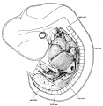
Fig 1. 11 mm embryo
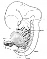
Fig 2. 17 mm embryo
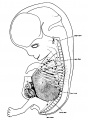
Fig 3. 31 mm embryo
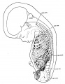
Fig 4. 65 mm embryo
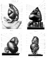
Fig. 5-8.
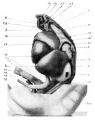
Fig 5. 11 mm embryo
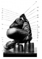
Fig 6. 17 mm embryo
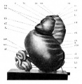
Fig 7. 31 mm embryo
Fig 8. 65 mm embryo


Reference
Jackson CM. On the developmental topography of the thoracic and abdominal viscera. (1909) Anat. Rec. 111: -396.
Cite this page: Hill, M.A. (2024, May 21) Embryology Jackson1909a fig04.jpg. Retrieved from https://embryology.med.unsw.edu.au/embryology/index.php/File:Jackson1909a_fig04.jpg
- © Dr Mark Hill 2024, UNSW Embryology ISBN: 978 0 7334 2609 4 - UNSW CRICOS Provider Code No. 00098G
File history
Click on a date/time to view the file as it appeared at that time.
| Date/Time | Thumbnail | Dimensions | User | Comment | |
|---|---|---|---|---|---|
| current | 11:09, 22 February 2018 | | 1,160 × 1,501 (187 KB) | Z8600021 (talk | contribs) | |
| 11:08, 22 February 2018 | 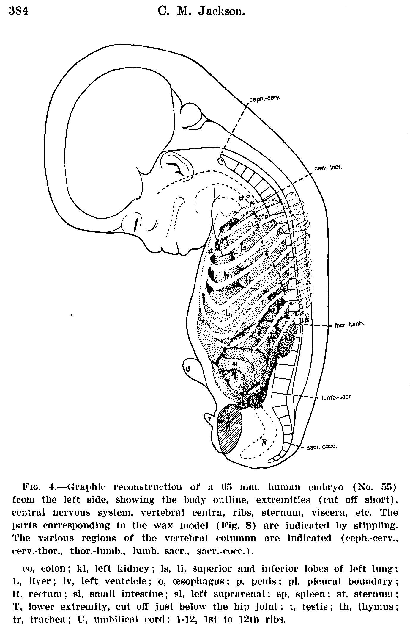 | 1,380 × 2,141 (327 KB) | Z8600021 (talk | contribs) | {{Jackson1909a figures}} |
You cannot overwrite this file.