File:Intestine histology 002.jpg: Difference between revisions
From Embryology
mNo edit summary |
mNo edit summary |
||
| Line 1: | Line 1: | ||
==Jejunum== | ==Jejunum== | ||
{{HE}} | {{HE}} [[:File:Intestine histology 001.jpg|Labeled Image]] | ||
Histology section of Jejunum | Histology section of Jejunum | ||
| Line 17: | Line 17: | ||
{{Intestine Histology}} | {{Intestine Histology}} | ||
{{Blue Histology}} | {{Blue Histology}} | ||
Latest revision as of 17:17, 2 March 2014
Jejunum
(Stain - Haematoxylin Eosin) Labeled Image
Histology section of Jejunum
The small intestine is divided into:
- Duodenum (25-30 cm)
- Jejunum (about first two-fifths of the rest)
- Ileum.
- The three segments merge imperceptibly and have the same basic histological organization.
- Accumulations of lymphocytes are common in the mucosa of the GIT, and they occur frequently in the small intestine.
- Lymphocytes can form very large aggregates in particular in the ileum where they may be covered by a specialised form of epithelium which facilitates their function in the immune-defence of the body against possible pathogens in the lumen of the intestine.
- These specialised parts of the small intestine are called Peyer's patches,
- Intestine Histology Links: Duodenum overview | Duodenum villi and crypts | Duodenum | Jejunum overview | Jejunum villus | Jejunum labeled | Jejunum unlabeled | Gastrointestinal Tract Histology | Intestine Development
Links: Histology | Histology Stains | Blue Histology images copyright Lutz Slomianka 1998-2009. The literary and artistic works on the original Blue Histology website may be reproduced, adapted, published and distributed for non-commercial purposes. See also the page Histology Stains.
Cite this page: Hill, M.A. (2024, May 8) Embryology Intestine histology 002.jpg. Retrieved from https://embryology.med.unsw.edu.au/embryology/index.php/File:Intestine_histology_002.jpg
- © Dr Mark Hill 2024, UNSW Embryology ISBN: 978 0 7334 2609 4 - UNSW CRICOS Provider Code No. 00098G
File history
Click on a date/time to view the file as it appeared at that time.
| Date/Time | Thumbnail | Dimensions | User | Comment | |
|---|---|---|---|---|---|
| current | 15:52, 23 February 2012 | 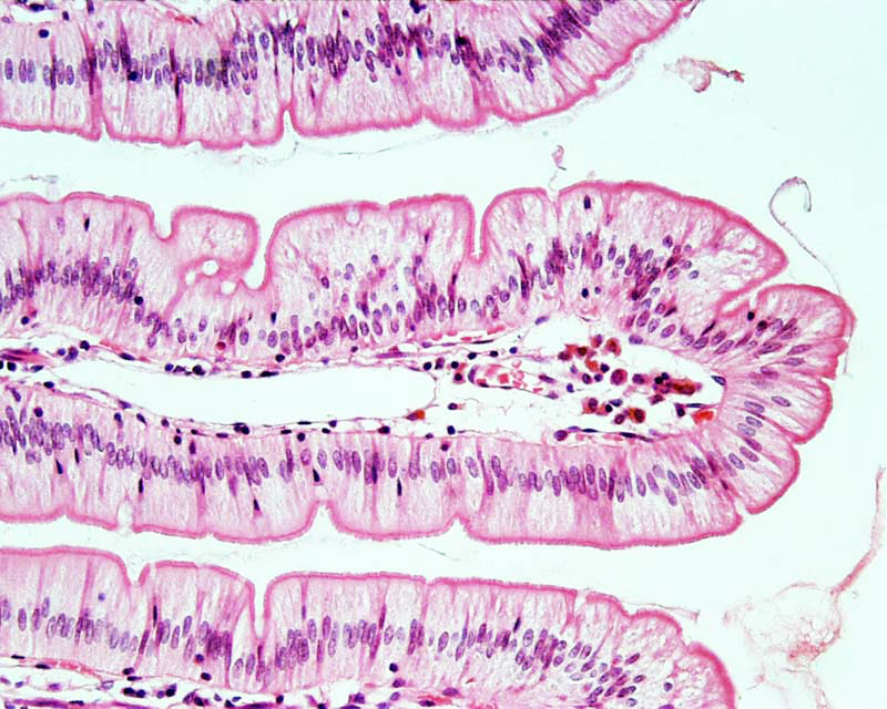 | 800 × 640 (130 KB) | Z8600021 (talk | contribs) | |
| 15:48, 23 February 2012 |  | 800 × 640 (83 KB) | Z8600021 (talk | contribs) | ==Jejunum== Histology section of Jejunum The small intestine is divided into duodenum (25-30 cm), jejunum (about first two-fifths of the rest) and ileum. The three segments merge imperceptibly and have the same basic histological organization. Accumula |
You cannot overwrite this file.
File usage
The following 4 pages use this file: