File:Hydatidiform mole 01.jpg: Difference between revisions
From Embryology
mNo edit summary |
mNo edit summary |
||
| Line 19: | Line 19: | ||
Figure 2: | Figure 2: | ||
[[Category:Human]] [[Category:Abnormal Development]] [[Category:Uterus]] | [[Category:Human]] [[Category:Abnormal Development]] [[Category:Uterus]][[Category:Hydatidiform Mole]] | ||
Latest revision as of 23:25, 11 May 2014
Hydatidiform Mole
Pathology specimen of entire gross uterus and molar component within the lower uterine segment extending through the internal cervical os.
Normal placental bed visualized within the fundal portion of the endometrial cavity. Multiple fibroids seen within the uterus.
- Links: Hydatidiform Mole
Reference
<pubmed>22928132</pubmed>| PMC3424659/ | Case Rep Obstet Gynecol.
Copyright
© 2012 Marijo Aguilera et al. This is an open access article distributed under the Creative Commons Attribution License, which permits unrestricted use, distribution, and reproduction in any medium, provided the original work is properly cited.
Figure 2:
File history
Click on a date/time to view the file as it appeared at that time.
| Date/Time | Thumbnail | Dimensions | User | Comment | |
|---|---|---|---|---|---|
| current | 22:14, 10 June 2013 | 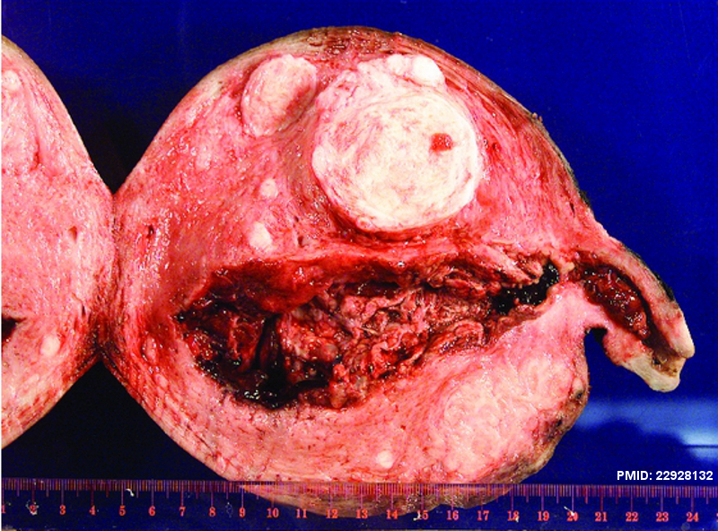 | 800 × 592 (634 KB) | Z8600021 (talk | contribs) | Figure 2: Pathology specimen of entire gross uterus and molar component within the lower uterine segment extending through the internal cervical os. Normal placental bed visualized within the fundal portion of the endometrial cavity. Multiple fibroids ... |
You cannot overwrite this file.
File usage
The following page uses this file: