File:Hunter1935 text-fig06.jpg
From Embryology

Size of this preview: 380 × 599 pixels. Other resolution: 731 × 1,153 pixels.
Original file (731 × 1,153 pixels, file size: 167 KB, MIME type: image/jpeg)
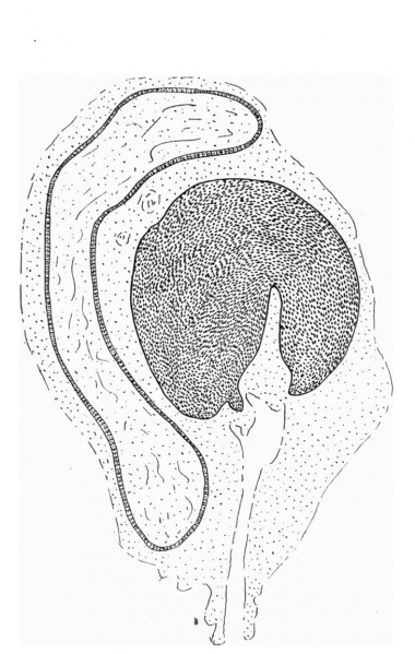
Text-Fig. 6. C.S. Glans region of penis from human foetus 100 mm. C.R. length
Note the two sub-glans folds closing off a portion of the desquamating epithelium within the developing glans portion of the urethra.
Reference
Hunter RH. Notes on the development of the prepuce. (1935) J Anat. 70: 68-75. PMID 17104576
Cite this page: Hill, M.A. (2024, April 28) Embryology Hunter1935 text-fig06.jpg. Retrieved from https://embryology.med.unsw.edu.au/embryology/index.php/File:Hunter1935_text-fig06.jpg
- © Dr Mark Hill 2024, UNSW Embryology ISBN: 978 0 7334 2609 4 - UNSW CRICOS Provider Code No. 00098G
File history
Click on a date/time to view the file as it appeared at that time.
| Date/Time | Thumbnail | Dimensions | User | Comment | |
|---|---|---|---|---|---|
| current | 16:30, 4 January 2017 | | 731 × 1,153 (167 KB) | Z8600021 (talk | contribs) |
You cannot overwrite this file.
File usage
The following page uses this file: