File:Humphrey1940 fig02.jpg

Original file (1,000 × 480 pixels, file size: 61 KB, MIME type: image/jpeg)
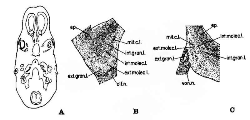
Fig. 2. Developing olfactory and accessory olfactory formations 26 mm fetus
Illustrations to show the position and morphology of the developing olfactory and accessory olfactory formations in the 26 mm. fetus. Erythrosin and toluidin blue preparation.
A, a horizontal section through the forebrain to indicate the plane of the sections and the location of the areas enlarged in B and C. X 3.
B, an enlargement of the region shown within the dotted lines on the left side of figure A, but at a level 80;; inferior to that of A. This drawing illustrates the beginning of lamination in the olfactory formation. X 30.
C, the area enclosed in the dotted lines on the right side of figure A, enlarged to illustrate the beginning of lamination in the accessory olfactory formation. X 30.
Reference
Humphrey T. The development of the olfactory and the accessory olfactory formations in human embryos and fetuses. (1940) J. Comp. Neurol. 431-468.
Cite this page: Hill, M.A. (2024, May 1) Embryology Humphrey1940 fig02.jpg. Retrieved from https://embryology.med.unsw.edu.au/embryology/index.php/File:Humphrey1940_fig02.jpg
- © Dr Mark Hill 2024, UNSW Embryology ISBN: 978 0 7334 2609 4 - UNSW CRICOS Provider Code No. 00098G
File history
Click on a date/time to view the file as it appeared at that time.
| Date/Time | Thumbnail | Dimensions | User | Comment | |
|---|---|---|---|---|---|
| current | 17:39, 24 October 2017 |  | 1,000 × 480 (61 KB) | Z8600021 (talk | contribs) | |
| 17:37, 24 October 2017 |  | 1,333 × 921 (198 KB) | Z8600021 (talk | contribs) | {{Ref-Humphrey1940}} |
You cannot overwrite this file.
File usage
The following page uses this file: