File:Human week 10 fetus 01.jpg: Difference between revisions
mNo edit summary |
mNo edit summary |
||
| (4 intermediate revisions by the same user not shown) | |||
| Line 1: | Line 1: | ||
==Human Female Fetus (10 week)== | ==Human Female Fetus (10 week)== | ||
Large image version of plane D, close to midline | Large image version of sagittal section (plane D), a sagittal plane close to midline {{HE}} 3 mm scale bar. | ||
The excerpted regions, shown in the table below, have further detailed descriptions of the structures and features. The headings for each link to the starting page for that system. Some of the excerpt images also include labels. | |||
{{Human Female Fetus Week 10 gallery}} | {{Human Female Fetus Week 10 gallery}} | ||
Latest revision as of 11:57, 30 May 2016
Human Female Fetus (10 week)
Large image version of sagittal section (plane D), a sagittal plane close to midline (Stain - Haematoxylin Eosin) 3 mm scale bar.
The excerpted regions, shown in the table below, have further detailed descriptions of the structures and features. The headings for each link to the starting page for that system. Some of the excerpt images also include labels.
- Human Female Fetus (week 10)
Sagittal Section (plane D)

Pituitary and Lamina Terminalis

Olfactory Nerve

Atlas and Axis

Sacrum

Oral Cavity

Epiglottis

Heart

Spleen

Midgut Herniation

Midgut Herniation (label)

Pelvic Region

Pelvic Region (label)


Related Images
Fetus (week 10) Planes A (most lateral), B (lateral), C (medial) and D (midline) from lateral towards the midline.
- Human Fetus - most lateral | lateral | medial | midline
- Head - most lateral | lateral | medial | midline
- Cerebellum - most lateral | lateral | medial | midline
- Urogenital Unlabelled - most lateral | lateral | medial | midline
- Urogenital Labelled - most lateral | lateral | medial | midline
- Large Images - midline
- Image Source: UNSW Embryology, no reproduction without permission.
Cite this page: Hill, M.A. (2024, May 7) Embryology Human week 10 fetus 01.jpg. Retrieved from https://embryology.med.unsw.edu.au/embryology/index.php/File:Human_week_10_fetus_01.jpg
- © Dr Mark Hill 2024, UNSW Embryology ISBN: 978 0 7334 2609 4 - UNSW CRICOS Provider Code No. 00098G
File history
Click on a date/time to view the file as it appeared at that time.
| Date/Time | Thumbnail | Dimensions | User | Comment | |
|---|---|---|---|---|---|
| current | 15:55, 17 June 2012 | 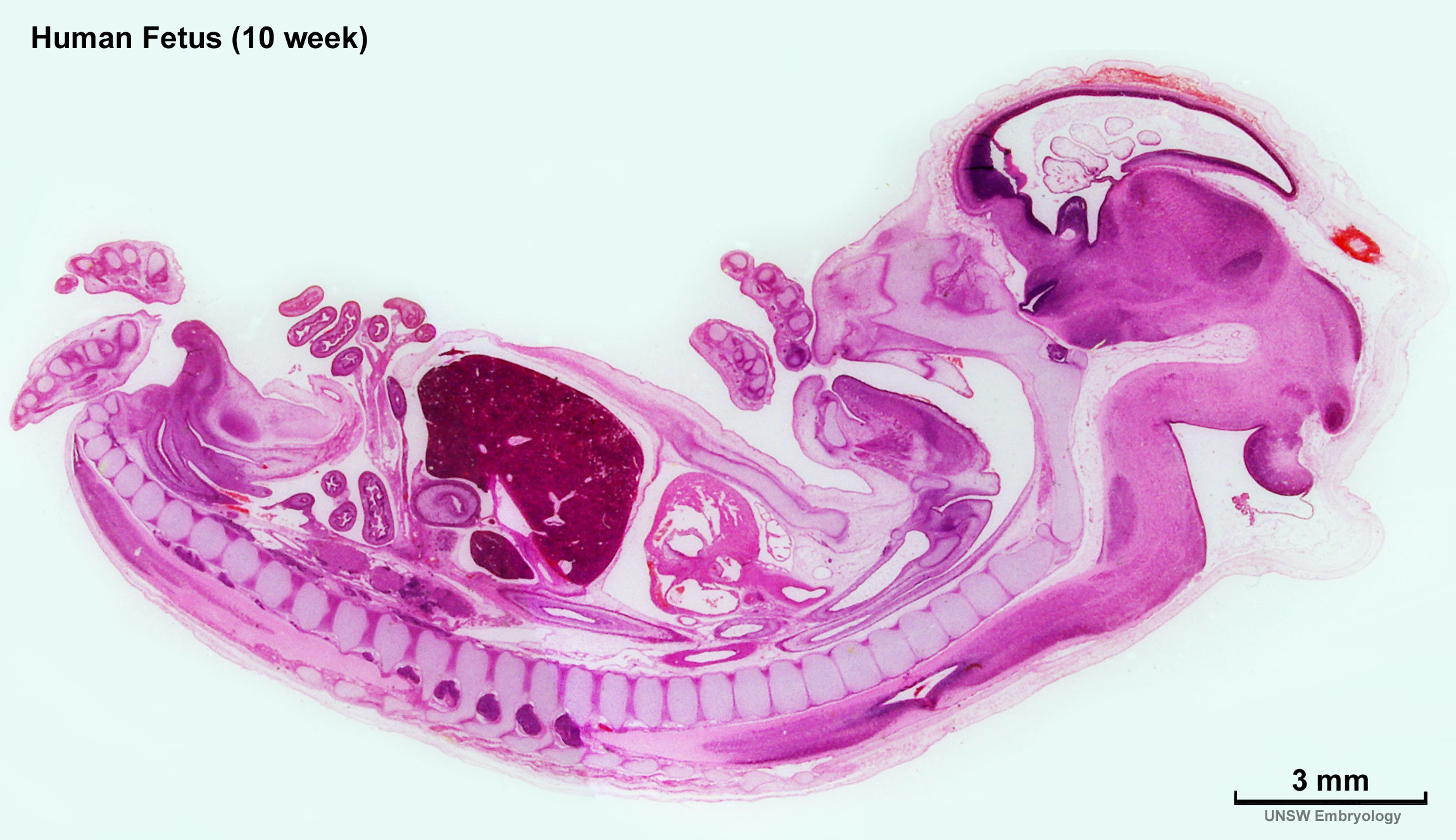 | 2,300 × 1,327 (448 KB) | Z8600021 (talk | contribs) |
You cannot overwrite this file.
File usage
The following 22 pages use this file:
- ANAT2341 Lab 11 - Embryo to Fetus
- BGDA Lecture - Development of the Nervous System
- BGDA Practical 12 - Embryo to Fetus
- BGDB Gastrointestinal - Activity 2
- BGDB Gastrointestinal - Fetal
- BGDB Sexual Differentiation - Fetal
- Fetal Development - 10 Weeks
- Foundations Practical - Week 9 to 36
- File:Human week 10 fetus 01.jpg
- File:Human week 10 fetus 03.jpg
- File:Human week 10 fetus 04.jpg
- File:Human week 10 fetus 05.jpg
- File:Human week 10 fetus 06.jpg
- File:Human week 10 fetus 07.jpg
- File:Human week 10 fetus 08.jpg
- File:Human week 10 fetus 09.jpg
- File:Human week 10 fetus 10.jpg
- File:Human week 10 fetus 11.jpg
- File:Human week 10 fetus 12.jpg
- File:Human week 10 fetus 23.jpg
- File:Human week 10 fetus 26.jpg
- Template:Human Female Fetus Week 10 gallery