File:Human sperm pathology EM02.jpg
Human_sperm_pathology_EM02.jpg (800 × 256 pixels, file size: 22 KB, MIME type: image/jpeg)
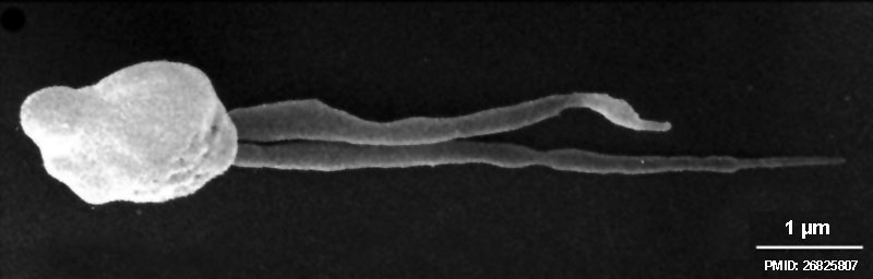
Human spermatozoa pathology - Two tails (EM)
Scanning electron micrograph of a dysplasia of the fibrous sheath (DFS) in human spermatozoa. Note the two thick, irregular and very short tails (length less than 10 μm, normal 50–60 μm).
Reference
<pubmed>26825807</pubmed>
Copyright
© The Author(s) 2016
Open Access - This article is distributed under the terms of the Creative Commons Attribution 4.0 International License (http://creativecommons.org/licenses/by/4.0/), which permits unrestricted use, distribution, and reproduction in any medium, provided you give appropriate credit to the original author(s) and the source, provide a link to the Creative Commons license, and indicate if changes were made.
Fig. 7 10815_2016_652_Fig7_HTML.jpg Original image panel (a) cropped from full figure, resized and labelled with PMID and scale bare size.
Cite this page: Hill, M.A. (2024, April 30) Embryology Human sperm pathology EM02.jpg. Retrieved from https://embryology.med.unsw.edu.au/embryology/index.php/File:Human_sperm_pathology_EM02.jpg
- © Dr Mark Hill 2024, UNSW Embryology ISBN: 978 0 7334 2609 4 - UNSW CRICOS Provider Code No. 00098G
File history
Click on a date/time to view the file as it appeared at that time.
| Date/Time | Thumbnail | Dimensions | User | Comment | |
|---|---|---|---|---|---|
| current | 11:55, 25 January 2017 | 800 × 256 (22 KB) | Z8600021 (talk | contribs) | ==Electron microscopic analysis of human sperm pathologies== '''a''' Scanning electron micrograph of a dysplasia of the fibrous sheath (DFS) in human spermatozoa. Note the two thick, irregular and very short tails (length less than 10 μm, normal 50�... |
You cannot overwrite this file.
File usage
There are no pages that use this file.