File:Human right ovary and tube 1.jpg
From Embryology
Size of this preview: 800 × 594 pixels. Other resolution: 916 × 680 pixels.
Original file (916 × 680 pixels, file size: 32 KB, MIME type: image/jpeg)
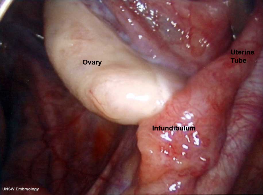
Human right ovary and tube as viewed by laparoscopy
Note the relative size and position of the ovary with respect to the uterine tube (fallopian tube). In the background the associated mesenteries and peritoneal cavity can be seen.
Image source: UNSW Embryology, no reproduction without permission.
File history
Click on a date/time to view the file as it appeared at that time.
| Date/Time | Thumbnail | Dimensions | User | Comment | |
|---|---|---|---|---|---|
| current | 09:31, 9 April 2010 | | 916 × 680 (32 KB) | S8600021 (talk | contribs) | Human right ovary and tube as viewed by laparoscopy == Image version links == Large 1000px | 800px | Medium 600px | [[:F |
You cannot overwrite this file.