File:Human placenta vascular CT 01.jpg
Original file (938 × 1,000 pixels, file size: 126 KB, MIME type: image/jpeg)
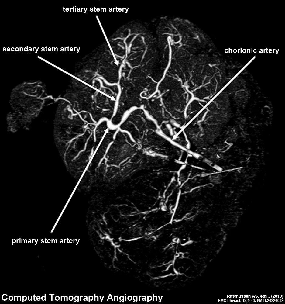
Human Placenta Vascular Bed - Computed tomography angiography (CTA)
Placenta is viewed from the fetal side of the placenta using CT imaging.
- Vascular Bed Links: Image - MRA and CTA | Image - magnetic resonance | Image - magnetic resonance labeled | Image - computed tomography | Placenta - Vascular Beds | Placenta Development
| Methodology technical information |
|---|
3D reconstruction made using Osirix software viewed from the fetal side of the placenta.
(text from original reference) |
Reference
Rasmussen AS, Lauridsen H, Laustsen C, Jensen BG, Pedersen SF, Uhrenholt L, Boel LW, Uldbjerg N, Wang T & Pedersen M. (2010). High-resolution ex vivo magnetic resonance angiography: a feasibility study on biological and medical tissues. BMC Physiol. , 10, 3. PMID: 20226038 DOI.
Copyright
© 2010 Rasmussen et al; licensee BioMed Central Ltd. This is an Open Access article distributed under the terms of the Creative Commons Attribution License (http://creativecommons.org/licenses/by/2.0), which permits unrestricted use, distribution, and reproduction in any medium, provided the original work is properly cited.
Original file name: Figure 4. 1472-6793-10-3-4-l.jpg (original figure has been cropped, resized and relabeled)
Cite this page: Hill, M.A. (2024, April 27) Embryology Human placenta vascular CT 01.jpg. Retrieved from https://embryology.med.unsw.edu.au/embryology/index.php/File:Human_placenta_vascular_CT_01.jpg
- © Dr Mark Hill 2024, UNSW Embryology ISBN: 978 0 7334 2609 4 - UNSW CRICOS Provider Code No. 00098G
File history
Click on a date/time to view the file as it appeared at that time.
| Date/Time | Thumbnail | Dimensions | User | Comment | |
|---|---|---|---|---|---|
| current | 13:35, 18 May 2013 | | 938 × 1,000 (126 KB) | Z8600021 (talk | contribs) |
You cannot overwrite this file.
File usage
The following 4 pages use this file: