File:Human ovary follicles light and electron microscopy 01.jpg

Original file (586 × 1,080 pixels, file size: 225 KB, MIME type: image/jpeg)
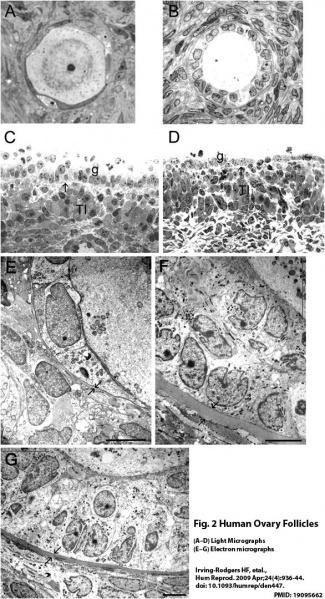
Light (A–D) and electron micrographs (E–G) of follicles from normal human ovaries.
Healthy primordial (A), a primary (B) follicle and a healthy 4 mm (C) and an atretic 5 mm (D) follicle. Ultrastructure of the follicle wall of primordial (E) and pre-antral follicles (F, G) Arrows indicate the position of the follicular basal lamina. G, membrana granulosa; T, theca interna; O, oocyte. Scale bar = 5 µm.
Reference
http://humrep.oxfordjournals.org/content/24/4/936.full
Copyright
© The Author 2008. Published by Oxford University Press on behalf of the European Society of Human Reproduction and Embryology. All rights reserved. For Permissions, please email: journals.permissions@oxfordjournals.org The online version of this article has been published under an open access model. Users are entitled to use, reproduce, disseminate, or display the open access version of this article for non-commercial purposes provided that: the original authorship is properly and fully attributed: the Journal and Oxford University Press are attributed as the original place of publication with the correct citation details given: if an article is subsequently reproduced or disseminated not in its entirety but only in part or as a derivative word this must be clearly indicated. For commercial re-use, please contact journals.permissions@oxfordjournals.org
Figure 2 has been extracted from original publication and relabelled with figure and reference information.
File history
Click on a date/time to view the file as it appeared at that time.
| Date/Time | Thumbnail | Dimensions | User | Comment | |
|---|---|---|---|---|---|
| current | 13:18, 30 April 2015 | | 586 × 1,080 (225 KB) | Z8600021 (talk | contribs) | Light (A–D) and electron micrographs (E–G) of follicles from normal human ovaries. Healthy primordial (A), a primary (B) follicle and a healthy 4 mm (C) and an atretic 5 mm (D) follicle. Ultrastructure of the follicle wall of primordial (E) and pr... |
You cannot overwrite this file.
File usage
The following page uses this file: