File:Human embryo neck 01.jpg

Original file (534 × 827 pixels, file size: 186 KB, MIME type: image/jpeg)
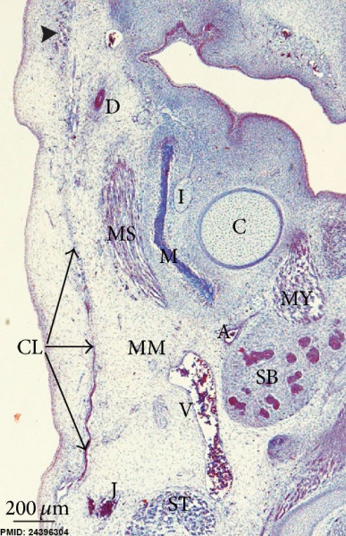
Human Embryo Neck Region
Human embryo GI4 (26.5 mm GL; 8 weeks of development). Frontal section. Azocarmine staining. The cervical lamina (CL) extends towards the mandibular region enclosing the masseter muscle (MS). Arrowhead: infraorbital lamina.
Bar: 200 μm.
A: Facial artery B: Buccinator muscle BB: Buccal branches of the facial nerve BF: Anlage of the buccal fat pad CP: Capsula propria of the parotid gland C: Meckel’s cartilage CL: Cervical lamina D: Parotid duct DA: Depressor anguli oris muscle DE: Dermis DF: Deep layer of the temporal fascia EP: Epidermis FN: Facial nerve G: Submandibular node lymph I: Inferior alveolar nerve IN: Infraorbital nerve J: External jugular vein L: Lingual nerve M: Mandible MM: Marginal mandibular branch of the facial nerve MS: Masseter muscle MY: Mylohyoid muscle P: Anlage of the parotid gland PL: Platysma muscle SA: Superficial adipose layer SB: Submandibular gland SF: Superficial layer of the temporal fascia ST: Sternocleidomastoid muscle T: Temporalis muscle TA: Superficial temporal artery TE: Temporalis muscle TF: Temporalis branches of the facial nerve TL: Temporal lamina V: Facial vein Z: Zygomaticus major muscle ZP: Zygomatic process of the squamous part of the temporal bone.
File history
Click on a date/time to view the file as it appeared at that time.
| Date/Time | Thumbnail | Dimensions | User | Comment | |
|---|---|---|---|---|---|
| current | 11:46, 28 March 2014 | | 534 × 827 (186 KB) | Z8600021 (talk | contribs) | ==Human Embryo Neck Region== Human embryo GI4 (26.5 mm GL; 8 weeks of development). Frontal section. Azocarmine staining. The cervical lamina (CL) extends towards the mandibular region enclosing the masseter muscle (MS). Arrowhead: infraorbital lami... |
You cannot overwrite this file.
File usage
There are no pages that use this file.