File:Human cochlea fetal development cartoon.jpg
Original file (592 × 1,200 pixels, file size: 96 KB, MIME type: image/jpeg)
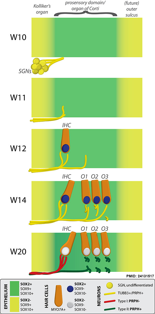
Human Cochlea Fetal Development Cartoon
Schematic diagram of neurosensory development in the basal turn of the human fetal cochlea by Gestational Week GA
|
Gestational Week GA
|
Abbreviations
|
Reference
Locher H, Frijns JH, van Iperen L, de Groot JC, Huisman MA & Chuva de Sousa Lopes SM. (2013). Neurosensory development and cell fate determination in the human cochlea. Neural Dev , 8, 20. PMID: 24131517 DOI.
Copyright
© 2013 Locher et al.; licensee BioMed Central Ltd. This is an open access article distributed under the terms of the Creative Commons Attribution License (http://creativecommons.org/licenses/by/2.0), which permits unrestricted use, distribution, and reproduction in any medium, provided the original work is properly cited.
Figure 9. 1749-8104-8-20-9.jpg Locher et al. Neural Development 2013 8:20 doi:10.1186/1749-8104-8-20 Original figure altered in size and labelling.
Cite this page: Hill, M.A. (2024, April 27) Embryology Human cochlea fetal development cartoon.jpg. Retrieved from https://embryology.med.unsw.edu.au/embryology/index.php/File:Human_cochlea_fetal_development_cartoon.jpg
- © Dr Mark Hill 2024, UNSW Embryology ISBN: 978 0 7334 2609 4 - UNSW CRICOS Provider Code No. 00098G
File history
Click on a date/time to view the file as it appeared at that time.
| Date/Time | Thumbnail | Dimensions | User | Comment | |
|---|---|---|---|---|---|
| current | 11:41, 29 June 2014 | | 592 × 1,200 (96 KB) | Z8600021 (talk | contribs) | Human cochlea fetal development cartoon © 2013 Locher et al.; licensee BioMed Central Ltd. This is an open access article distributed under the terms of the Creative Commons Attribution License (http://creativecommons.org/licenses/by/2.0), which per... |
You cannot overwrite this file.
File usage
The following page uses this file: