File:Human axial skeleton- axis development 01.jpg: Difference between revisions
No edit summary |
No edit summary |
||
| Line 2: | Line 2: | ||
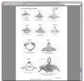
This is a plate from a historic study of the human axis vertebra. | This is a plate from a historic study of the human axis vertebra. | ||
PLATE IX. | PLATE IX. | ||
Fig.1.Upper surface of axis at 11th week x5. a, cartilaginous body; b, bony rod in arch. | '''Fig. 1.''' Upper surface of axis at 11th week x5. a, cartilaginous body; b, bony rod in arch. | ||
Fig.2.Coronal section of axis at 16th week x2. c, dens; d, bony nucleus in body; other letters as last. | '''Fig. 2.''' Coronal section of axis at 16th week x2. c, dens; d, bony nucleus in body; other letters as last. | ||
Fig.3.Coronal section of axis at 19th week. d, united body nuclei; e, densnuclei. | '''Fig. 3.''' Coronal section of axis at 19th week. d', united body nuclei; e, densnuclei. | ||
Fig. 4 | '''Fig. 4.''' Similar section at 22nd week. | ||
Fig. 5. Unusual case of delayed union of lateral dens centres in infant of 5 months old. | '''Fig. 5.''' Unusual case of delayed union of lateral dens centres in infant of 5 months old. | ||
Fig.6. Axis at birth. e', united dens centres. | '''Fig. 6.''' Axis at birth. e', united dens centres. | ||
Fig.7. Axis of child 15 months old, seen from below. f, hypo- chordal nucleus. | '''Fig. 7.''' Axis of child 15 months old, seen from below. f, hypo- chordal nucleus. | ||
Fig. 8. Coronal section of axis of 28 months child, showing hypo- chordalepiphysis. | '''Fig. 8.''' Coronal section of axis of 28 months child, showing hypo- chordalepiphysis. | ||
Fig. 9. Axis of child 45 months old, showing the apical epiphysis, g,ofthedens. | '''Fig. 9.''' Axis of child 45 months old, showing the apical epiphysis, g,ofthedens. | ||
Fig. 10. Coronal section of adult axis, showing, h, the lenticular cartilage between the dens and the body of the axis; and, i, the inferiorepiphysisofthebody. | '''Fig. 10.''' Coronal section of adult axis, showing, h, the lenticular cartilage between the dens and the body of the axis; and, i, the inferiorepiphysisofthebody. | ||
==Reference== | ==Reference== | ||
Revision as of 08:54, 9 March 2011
Human Axis Development
This is a plate from a historic study of the human axis vertebra.
PLATE IX.
Fig. 1. Upper surface of axis at 11th week x5. a, cartilaginous body; b, bony rod in arch.
Fig. 2. Coronal section of axis at 16th week x2. c, dens; d, bony nucleus in body; other letters as last.
Fig. 3. Coronal section of axis at 19th week. d', united body nuclei; e, densnuclei.
Fig. 4. Similar section at 22nd week.
Fig. 5. Unusual case of delayed union of lateral dens centres in infant of 5 months old.
Fig. 6. Axis at birth. e', united dens centres.
Fig. 7. Axis of child 15 months old, seen from below. f, hypo- chordal nucleus.
Fig. 8. Coronal section of axis of 28 months child, showing hypo- chordalepiphysis.
Fig. 9. Axis of child 45 months old, showing the apical epiphysis, g,ofthedens.
Fig. 10. Coronal section of adult axis, showing, h, the lenticular cartilage between the dens and the body of the axis; and, i, the inferiorepiphysisofthebody.
Reference
<pubmed>17232080</pubmed>| PMC1328386
File history
Click on a date/time to view the file as it appeared at that time.
| Date/Time | Thumbnail | Dimensions | User | Comment | |
|---|---|---|---|---|---|
| current | 08:51, 9 March 2011 |  | 763 × 1,095 (95 KB) | S8600021 (talk | contribs) | |
| 08:49, 9 March 2011 |  | 1,248 × 1,200 (139 KB) | S8600021 (talk | contribs) | PLATE IX. Fig.1.Upper surface of axis at 11th week x5. a, cartilaginous body; b, bony rod in arch. Fig.2.Coronal section of axis at 16th week x2. c, dens; d, bony nucleus in body; other letters as last. Fig.3.Coronal section of axis at 19th week. d, u |
You cannot overwrite this file.
File usage
The following page uses this file: