File:Human- adult ovary epithelial cords and primary follicles.jpg
Human-_adult_ovary_epithelial_cords_and_primary_follicles.jpg (600 × 384 pixels, file size: 51 KB, MIME type: image/jpeg)
=Distribution of epithelial cords and primary follicles in the ovarian cortex
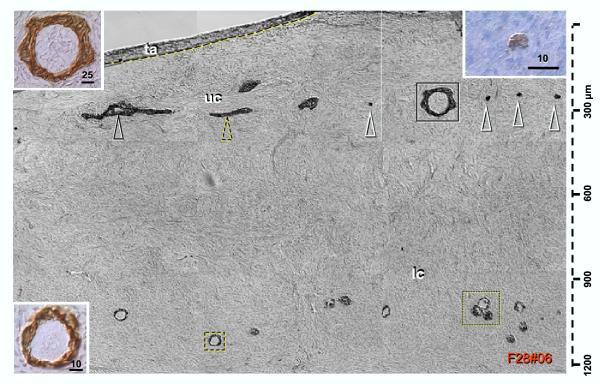
Panoramic view composed of nine images shows ovarian tunica albuginea filled with CK18+ mesenchymal cells, upper cortex (uc) with epithelial channels (black arrowhead), cords (dashed and white arrowheads – see right inset) and follicle-like structures (solid box, see upper left inset for details). Lower cortex (Ic) shows isolated (dashed box – see lower left inset) and grouped primary follicles (dotted box). Bars in insets indicate μm. Cytokeratin 18 immunostaining, hematoxylin counterstain.
This figure shows formation of epithelial channels (arrowhead), longitudinally (dashed arrowhead) and perpendicularly viewed solid epithelial cords (white arrowheads and right inset), and follicle like structures containing stromal elements (solid box, see left top inset for detail) in the upper cortex (uc). These structures were arranged in a surprisingly straight row (see also Fig. 1D). In some ovaries a similar orientation of primary follicles was observed in the lower ovarian cortex (Ic).
Original File Name: 1477-7827-2-20-3.jpg
Reference
Origin of germ cells and formation of new primary follicles in adult human ovaries. Bukovsky A, Caudle MR, Svetlikova M, Upadhyaya NB. Reprod Biol Endocrinol. 2004 Apr 28;2:20. PMID: 15115550 | Reprod Biol Endocrinol.
Bukovsky et al. Reproductive Biology and Endocrinology 2004 2:20 doi:10.1186/1477-7827-2-20 http://www.rbej.com/content/2/1/20
© 2004 Bukovsky et al; licensee BioMed Central Ltd. This is an Open Access article: verbatim copying and redistribution of this article are permitted in all media for any purpose, provided this notice is preserved along with the article's original URL.
File history
Click on a date/time to view the file as it appeared at that time.
| Date/Time | Thumbnail | Dimensions | User | Comment | |
|---|---|---|---|---|---|
| current | 02:00, 18 April 2010 |  | 600 × 384 (51 KB) | S8600021 (talk | contribs) | Distribution of epithelial cords and primary follicles in the ovarian cortex. Panoramic view composed of nine images shows ovarian tunica albuginea filled with CK18+ mesenchymal cells, upper cortex (uc) with epithelial channels (black arrowhead), cords |
You cannot overwrite this file.
File usage
There are no pages that use this file.
