File:Huber1915 1fig16.jpg
Huber1915_1fig16.jpg (800 × 416 pixels, file size: 50 KB, MIME type: image/jpeg)
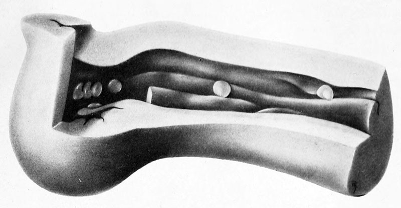
Fig. 16 Model of the segment of the rat oviduct
Rat No. 57, 3 days, 17 hours, containing the ova shown in fig. 15. X 50. A portion of the wall of the oviduct and a part of the major folds of the mucosa are removed in the drawing so as to expose the contained ova. The relative position of the seven ova found in the tube is shown, as also the extent and character of the folds of the mucosa. The exact form of each of the several ova could not be reproduced in the model at the magnification used; their position is given correctly.
| Historic Disclaimer - information about historic embryology pages |
|---|
| Pages where the terms "Historic" (textbooks, papers, people, recommendations) appear on this site, and sections within pages where this disclaimer appears, indicate that the content and scientific understanding are specific to the time of publication. This means that while some scientific descriptions are still accurate, the terminology and interpretation of the developmental mechanisms reflect the understanding at the time of original publication and those of the preceding periods, these terms, interpretations and recommendations may not reflect our current scientific understanding. (More? Embryology History | Historic Embryology Papers) |
- Albino Rat Links: Fig 14. Right Oviduct | Fig 15. 8 and 11-cell stages | The Development of the Albino Rat 1915
Cite this page: Hill, M.A. (2024, April 27) Embryology Huber1915 1fig16.jpg. Retrieved from https://embryology.med.unsw.edu.au/embryology/index.php/File:Huber1915_1fig16.jpg
- © Dr Mark Hill 2024, UNSW Embryology ISBN: 978 0 7334 2609 4 - UNSW CRICOS Provider Code No. 00098G
| Historic Disclaimer - information about historic embryology pages |
|---|
| Pages where the terms "Historic" (textbooks, papers, people, recommendations) appear on this site, and sections within pages where this disclaimer appears, indicate that the content and scientific understanding are specific to the time of publication. This means that while some scientific descriptions are still accurate, the terminology and interpretation of the developmental mechanisms reflect the understanding at the time of original publication and those of the preceding periods, these terms, interpretations and recommendations may not reflect our current scientific understanding. (More? Embryology History | Historic Embryology Papers) |
Cite this page: Hill, M.A. (2024, April 27) Embryology Huber1915 1fig16.jpg. Retrieved from https://embryology.med.unsw.edu.au/embryology/index.php/File:Huber1915_1fig16.jpg
- © Dr Mark Hill 2024, UNSW Embryology ISBN: 978 0 7334 2609 4 - UNSW CRICOS Provider Code No. 00098G
File history
Click on a date/time to view the file as it appeared at that time.
| Date/Time | Thumbnail | Dimensions | User | Comment | |
|---|---|---|---|---|---|
| current | 11:34, 7 April 2013 |  | 800 × 416 (50 KB) | Z8600021 (talk | contribs) | ==Fig. 16 Model of the segment of the rat oviduct== Rat No. 57, 3 days, 17 hours, containing the ova shown in fig. 15. X 50. A portion of the wall of the oviduct and a part of the major folds of the mucosa are removed in the drawing so as to expose th... |
You cannot overwrite this file.
File usage
The following page uses this file:

