File:Huber1915 1fig05.jpg

Huber1915_1fig05.jpg (800 × 561 pixels, file size: 84 KB, MIME type: image/jpeg)
Fig. 5 Camera lucida drawing of a portion of a section of the left oviduct of rat
Camera lucida drawing of a portion of a section of the left oviduct of rat No. 104, 24 hours, 15 rain. X 100. Three ova with a few discus cells, are shown as lying free within the lumen.
The ova are in the pronuclear stage, not shown in this section, but readily ascertained by tracing through the scries. The loop of the oviduct here shown in section is cut longitudinally, thus the folds of the mucosa are not prominent.
| Historic Disclaimer - information about historic embryology pages |
|---|
 |
- Albino Rat Links: Fig 14. Right Oviduct | Fig 15. 8 and 11-cell stages | The Development of the Albino Rat 1915
Cite this page: Hill, M.A. (2024, April 26) Embryology Huber1915 1fig05.jpg. Retrieved from https://embryology.med.unsw.edu.au/embryology/index.php/File:Huber1915_1fig05.jpg
- © Dr Mark Hill 2024, UNSW Embryology ISBN: 978 0 7334 2609 4 - UNSW CRICOS Provider Code No. 00098G
| Historic Disclaimer - information about historic embryology pages |
|---|
|
Cite this page: Hill, M.A. (2024, April 26) Embryology Huber1915 1fig05.jpg. Retrieved from https://embryology.med.unsw.edu.au/embryology/index.php/File:Huber1915_1fig05.jpg
- © Dr Mark Hill 2024, UNSW Embryology ISBN: 978 0 7334 2609 4 - UNSW CRICOS Provider Code No. 00098G
File history
Click on a date/time to view the file as it appeared at that time.
| Date/Time | Thumbnail | Dimensions | User | Comment | |
|---|---|---|---|---|---|
| current | 10:43, 7 April 2013 | | 800 × 561 (84 KB) | Z8600021 (talk | contribs) | |
| 00:23, 5 April 2013 | 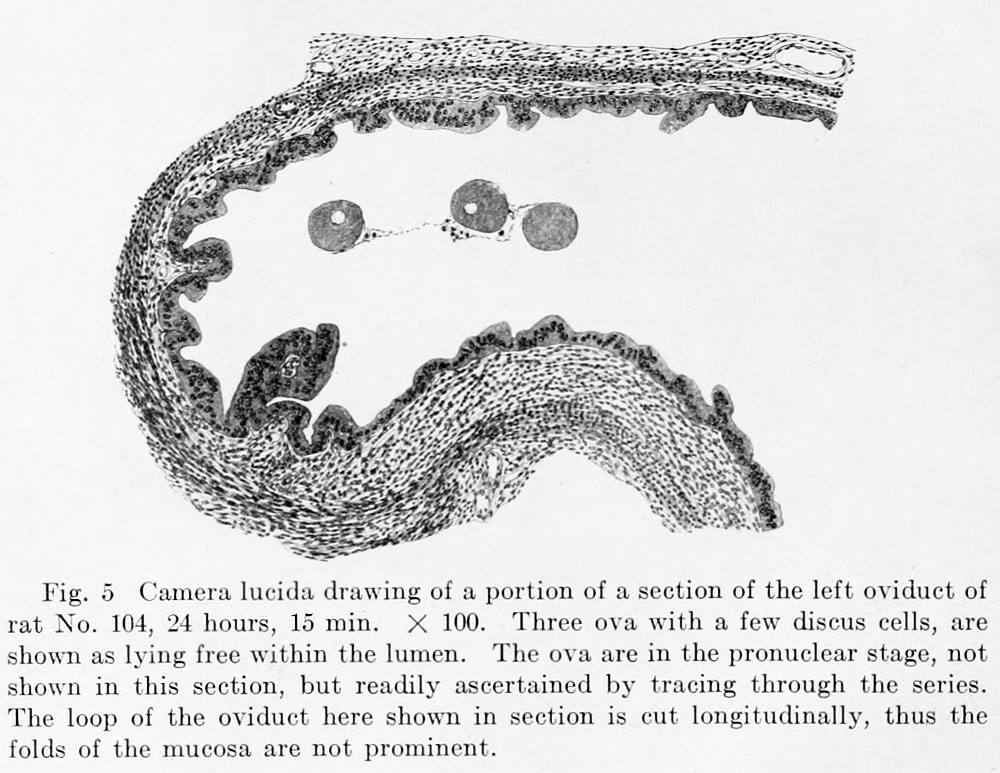 | 1,000 × 773 (139 KB) | Z8600021 (talk | contribs) | {{Huber1915 figures}} {{Huber1915_footer}} |
You cannot overwrite this file.
File usage
The following 2 pages use this file: