File:His1897 plate03.jpg
Original file (1,738 × 2,808 pixels, file size: 977 KB, MIME type: image/jpeg)
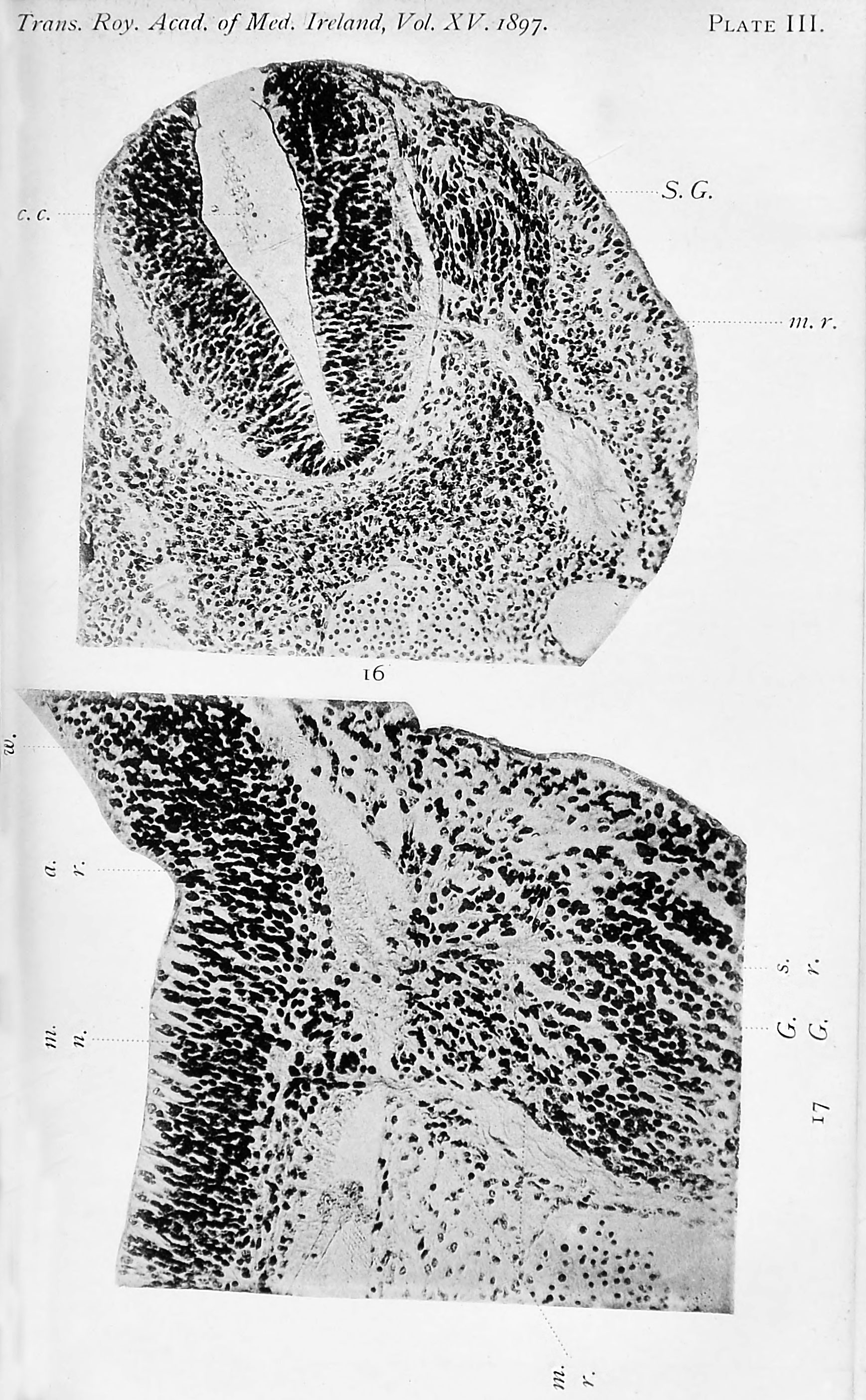
Plate III
Fig. 16. Transverse section through the spinal part of the neural tube of a human embryo of four weeks. It shows the early condition of the roots of a spinal nerve. A diagrammatic representation of this is given in Fig. 2 in the text.
c.c. Central canal of neural tube.
m.r. Motor fibres from the neuroblasts in the wall of the tube, growing out to form the anterior nerve-root.
S.G. Spinal ganglion, from the cells of which the fibres grow into the wall of the neural tube and thus form the posterior nerve-root.
Fig. 17. Section through a portion of the wall of the brain tube at the level of the origin of the fifth cranial nerve. It has been found necessary to turn this figure round on its side so as to make it fit into the plate.
w. Wall of the brain-tube.
G.G. Gasserian ganglion. x.r. Sensory root, formed of fibres growing out from the cells of the ganglion and entering the wall of the neural tube. a.T. The same fibres as represented by s.r. cut transversely owing to their having turned downwards to form the spinal root of the fifth nerve.
m.n. Neuroblasts of the motor nucleus of the fifth nerve giving origin to the motor fibres m.r . which are seen growing out from the neural wall. m.r. Fibres of the motor root of the fifth nerve.
Human embryo of four weeks.
Fig. 16. Transverse section through the spinal part of the neural tube of a human embryo of four weeks. It shows the early condition of the roots of a spinal nerve. A diagrammatic representation of this is given in Fig. 2 in the text (p. 5).
Fig. 17. Section through a portion of the wall of the brain tube at the level of the origin of the fifth cranial nerve. Jt has been found necessary to turn this figure round on its side so as to make it fit into the plate.


| Historic Disclaimer - information about historic embryology pages |
|---|
 |
- Links: Fig 1 | Fig 2 | Fig 3 | Fig 4 | Fig 5 | Fig 6 | Plate 1 | Fig 7 | Fig 8 | Fig 9 | Fig 10 | Fig 11 | Plate 2 | Fig 12 | Fig 13 | Fig 14 | Fig 15 | Plate 3 | Fig 16 | Fig 17 | Plate 4 | Fig 18 | Fig 19 | Fig 20 | Fig 21 | Fig 22 | Fig 23 | Plate 5 | Fig 24 | Fig 25 | Fig 26 | Fig 27 | Fig 28 | His 1897 | Wilhelm His | Historic Embryology Papers
Reference
His W. Address upon the development of the brain. (1897) Trans. Royal Acad. Medicine Ireland.
Cite this page: Hill, M.A. (2024, April 27) Embryology His1897 plate03.jpg. Retrieved from https://embryology.med.unsw.edu.au/embryology/index.php/File:His1897_plate03.jpg
- © Dr Mark Hill 2024, UNSW Embryology ISBN: 978 0 7334 2609 4 - UNSW CRICOS Provider Code No. 00098G
File history
Click on a date/time to view the file as it appeared at that time.
| Date/Time | Thumbnail | Dimensions | User | Comment | |
|---|---|---|---|---|---|
| current | 18:58, 17 January 2016 | | 1,738 × 2,808 (977 KB) | Z8600021 (talk | contribs) |
You cannot overwrite this file.
File usage
The following page uses this file: