File:His1897 plate02.jpg

Original file (1,767 × 2,989 pixels, file size: 895 KB, MIME type: image/jpeg)
Plate II
Fig. 12. Microphotograph of a portion of a transverse section through the whole thickness of the neural tube. The arrangement of the neuroblasts and of the spongioblasts is well seen. The limiting membrane on the inner aspect of the tube is uppermost in the figure. Human embryo of four weeks.
Fig. 13. Transverse section through the early medulla showing the rhombic lip. r.l. Rhombic lip. G.x. Ganglion of the vagus.
Fig. 14. Mesial section through the head of a human embryo of the seventh week. It shows the great expansion of the cavity of the brain-part of the neural tube as well as the strongly-marked brain flexures.
Fig. 15. Photograph of a human embryo of six weeks. The different subdivisions of the brain shining through the thin head-coverings are well seen, and the cervical, pontine and mid-brain flexures observed, in section, in Fig. 14 are prominently brought out. Compare with Fig. 6 in the text.
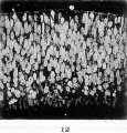
Fig. 12. Microphotograph of a portion of a transverse section through the whole thickness of the neural tube. The arrangement of the neuroblasts and of the spongioblasts is well seen. The limiting membrane on the inner aspect of the tube is uppermost in the figure. Human embryo of four weeks.
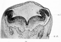
Fig. 13. Transverse section through the early medulla showing the rhombic lip. r.l. Rhombic lip. G.x. Ganglion of the vagus.
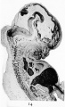
Fig. 14. Mesial section through the head of a human embryo of the seventh week. It shows the great expansion of the cavity of the brain-part of the neural tube as well as the strongly-marked brain flexures.


| Historic Disclaimer - information about historic embryology pages |
|---|
 |
- Links: Fig 1 | Fig 2 | Fig 3 | Fig 4 | Fig 5 | Fig 6 | Plate 1 | Fig 7 | Fig 8 | Fig 9 | Fig 10 | Fig 11 | Plate 2 | Fig 12 | Fig 13 | Fig 14 | Fig 15 | Plate 3 | Fig 16 | Fig 17 | Plate 4 | Fig 18 | Fig 19 | Fig 20 | Fig 21 | Fig 22 | Fig 23 | Plate 5 | Fig 24 | Fig 25 | Fig 26 | Fig 27 | Fig 28 | His 1897 | Wilhelm His | Historic Embryology Papers
Reference
His W. Address upon the development of the brain. (1897) Trans. Royal Acad. Medicine Ireland.
Cite this page: Hill, M.A. (2024, April 27) Embryology His1897 plate02.jpg. Retrieved from https://embryology.med.unsw.edu.au/embryology/index.php/File:His1897_plate02.jpg
- © Dr Mark Hill 2024, UNSW Embryology ISBN: 978 0 7334 2609 4 - UNSW CRICOS Provider Code No. 00098G
File history
Click on a date/time to view the file as it appeared at that time.
| Date/Time | Thumbnail | Dimensions | User | Comment | |
|---|---|---|---|---|---|
| current | 19:06, 17 January 2016 | | 1,767 × 2,989 (895 KB) | Z8600021 (talk | contribs) |
You cannot overwrite this file.
File usage
The following page uses this file: