File:His1897 fig01.jpg

Original file (768 × 1,000 pixels, file size: 127 KB, MIME type: image/jpeg)
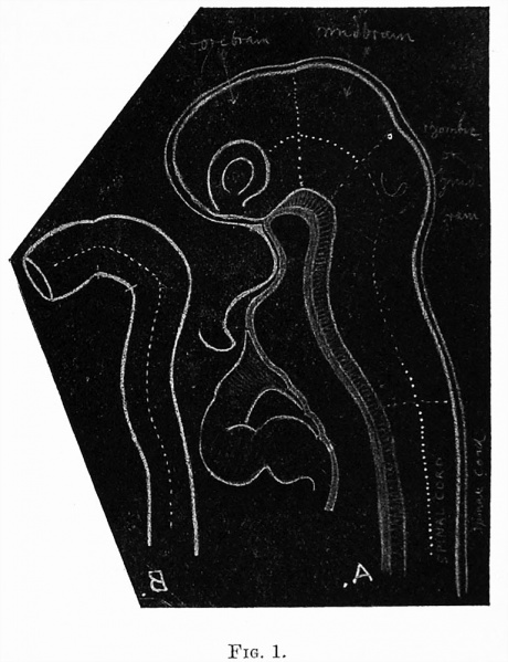
Fig. 1. Photographic reproduction of black-board drawing made by Prof. His for his Lecture
B. Diagrammatic representation of early condition of the neural tube, showing the manner in which it becomes bent on itself.
A. Diagrammatic representation of a mesial longitudinal section through the head and neck of a human embryo. The brain-flexures and the optic vesicle are seen, and the manner in which the fore-brain is bent around the anterior extremity of the notochord and foregut is indicated. The early heart is shown bulging out in front of the foregut. The different sub-divisions of the brain are marked off from each other by dotted lines.
In both diagrams (A as well as B) a dotted line running in the long axis of the neural tube indicates the separation of the alar from the basal lamina of the lateral wall.
| Historic Disclaimer - information about historic embryology pages |
|---|
 |
- Links: Fig 1 | Fig 2 | Fig 3 | Fig 4 | Fig 5 | Fig 6 | Plate 1 | Fig 7 | Fig 8 | Fig 9 | Fig 10 | Fig 11 | Plate 2 | Fig 12 | Fig 13 | Fig 14 | Fig 15 | Plate 3 | Fig 16 | Fig 17 | Plate 4 | Fig 18 | Fig 19 | Fig 20 | Fig 21 | Fig 22 | Fig 23 | Plate 5 | Fig 24 | Fig 25 | Fig 26 | Fig 27 | Fig 28 | His 1897 | Wilhelm His | Historic Embryology Papers
Reference
His W. Address upon the development of the brain. (1897) Trans. Royal Acad. Medicine Ireland.
Cite this page: Hill, M.A. (2024, April 27) Embryology His1897 fig01.jpg. Retrieved from https://embryology.med.unsw.edu.au/embryology/index.php/File:His1897_fig01.jpg
- © Dr Mark Hill 2024, UNSW Embryology ISBN: 978 0 7334 2609 4 - UNSW CRICOS Provider Code No. 00098G
File history
Click on a date/time to view the file as it appeared at that time.
| Date/Time | Thumbnail | Dimensions | User | Comment | |
|---|---|---|---|---|---|
| current | 10:12, 18 January 2016 | | 768 × 1,000 (127 KB) | Z8600021 (talk | contribs) | |
| 10:08, 18 January 2016 |  | 1,200 × 1,658 (290 KB) | Z8600021 (talk | contribs) |
You cannot overwrite this file.
File usage
The following page uses this file: