File:Hilfer1990 Fig10.jpg
Original file (1,200 × 863 pixels, file size: 77 KB, MIME type: image/jpeg)
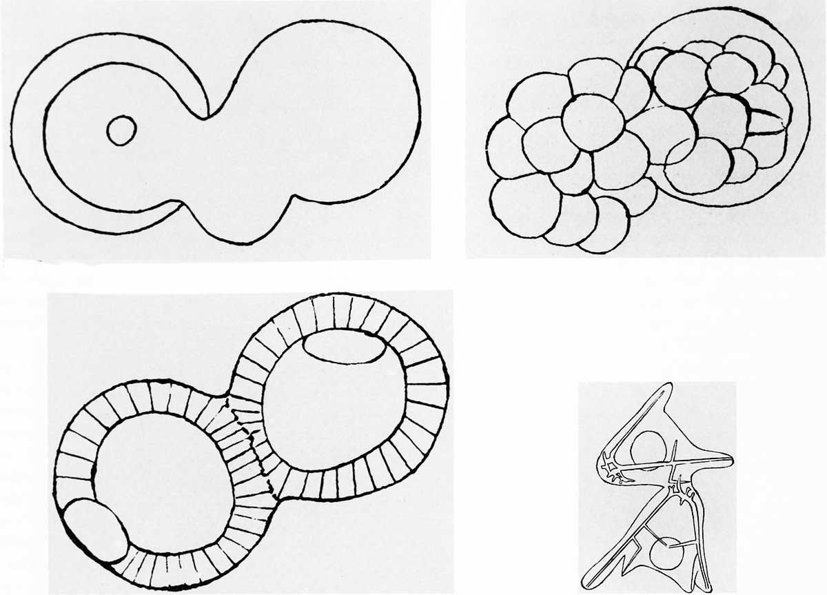
Figure 10. J. Loeb (1893) sea urchin eggs
Experiments by J. Loeb, as in this paper from 1893, gave additional support to the argument that sea urchin eggs do not contain specialized areas of cytoplasmic determinants. Treatment of fertilized eggs with hypotonic sea water resulted in the formation of cytoplasmic blebs (upper left) that remained attached to the main embryo during cleavage (upper right) and resulted in doubled embryos at the blastula stage (lower left). These embryos developed into Siamese twins (lower right). The egg cytoplasm was able to form most of the parts of more than one individual.
- Figures: Fig 1. by N. Hartsoeker 1694 | Fig 2. by M. Malpighi 1673 | Fig 3. by C.E. von Baer 1827 | Fig 4. by W. Roux 1888 | Fig 5. by H. Driesch 1892 | Fig 6. Louis Agassiz | Fig 7. Leonard W. Williams c1900 | Fig 8. by Conklin 1905 | Fig 9. by Wilson 1892 | Fig 10. by Loeb 1893 | Fig 11. by E. B. Wilson 1904 | Fig 12. by O.E. Schotte | Fig 13. by Spemann and H. Mangold 1924 | Fig 14. by S. Horstadius 1928 | Fig 15. by R. G. Harrison 1921 | Fig 16. by Townes and Holtfreter 1955
Cite this page: Hill, M.A. (2024, April 27) Embryology Hilfer1990 Fig10.jpg. Retrieved from https://embryology.med.unsw.edu.au/embryology/index.php/File:Hilfer1990_Fig10.jpg
- © Dr Mark Hill 2024, UNSW Embryology ISBN: 978 0 7334 2609 4 - UNSW CRICOS Provider Code No. 00098G
File history
Click on a date/time to view the file as it appeared at that time.
| Date/Time | Thumbnail | Dimensions | User | Comment | |
|---|---|---|---|---|---|
| current | 10:05, 28 August 2014 | | 1,200 × 863 (77 KB) | Z8600021 (talk | contribs) | {{Hilfer1990figures}} |
You cannot overwrite this file.
File usage
The following page uses this file: