File:Hilfer1990 Fig08.jpg
Original file (1,498 × 2,000 pixels, file size: 373 KB, MIME type: image/jpeg)
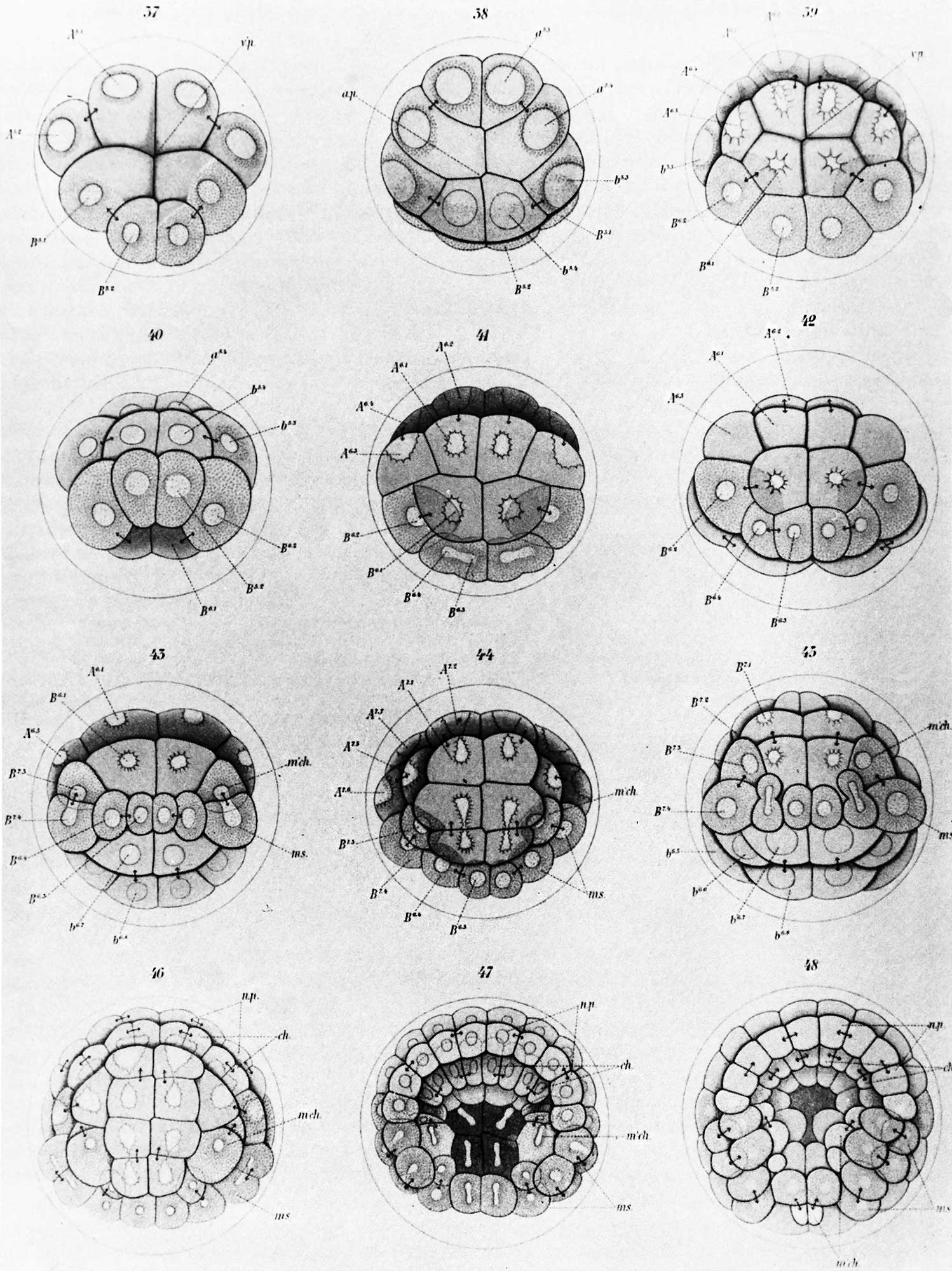
Figure 8. Conklin (1905) early cleavage stages of the tunicate
Diagrams of early cleavage stages of the tunicate, Cynthia partita, from Conklin (1905). The four cells at the bottom of the upper left figure are traced through succeeding cleavages into a series of blastomeres that give rise to segmental muscles and other mesodermal derivatives. This is an example of the painstaking care that was needed to trace regionalization of egg cytoplasm through development.
tunicate - (sea squirt) a marine invertebrate animal (subphylum Tunicata, phylum Chordata) includes all animals with dorsal nerve cords and notochords.
- Figures: Fig 1. by N. Hartsoeker 1694 | Fig 2. by M. Malpighi 1673 | Fig 3. by C.E. von Baer 1827 | Fig 4. by W. Roux 1888 | Fig 5. by H. Driesch 1892 | Fig 6. Louis Agassiz | Fig 7. Leonard W. Williams c1900 | Fig 8. by Conklin 1905 | Fig 9. by Wilson 1892 | Fig 10. by Loeb 1893 | Fig 11. by E. B. Wilson 1904 | Fig 12. by O.E. Schotte | Fig 13. by Spemann and H. Mangold 1924 | Fig 14. by S. Horstadius 1928 | Fig 15. by R. G. Harrison 1921 | Fig 16. by Townes and Holtfreter 1955
Cite this page: Hill, M.A. (2024, April 26) Embryology Hilfer1990 Fig08.jpg. Retrieved from https://embryology.med.unsw.edu.au/embryology/index.php/File:Hilfer1990_Fig08.jpg
- © Dr Mark Hill 2024, UNSW Embryology ISBN: 978 0 7334 2609 4 - UNSW CRICOS Provider Code No. 00098G
File history
Click on a date/time to view the file as it appeared at that time.
| Date/Time | Thumbnail | Dimensions | User | Comment | |
|---|---|---|---|---|---|
| current | 09:50, 28 August 2014 | | 1,498 × 2,000 (373 KB) | Z8600021 (talk | contribs) |
You cannot overwrite this file.
File usage
The following page uses this file: