File:Hilfer1990 Fig04.jpg

Original file (1,855 × 2,000 pixels, file size: 238 KB, MIME type: image/jpeg)
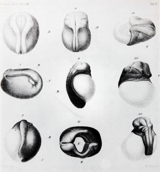
Figure 4. W. Roux Surface views of frog embryos
Surface views of frog embryos from W. Roux (1888). His Figures 5 and 6 show normal embryos at early and late stages of formation of the neural tube. His Figures 7 to 10 show embryos in which one blastomere was killed at the two-cell stage. In Figure 7, the ectoderm has covered much of the surface of the dead blastomere whereas much less repair has occurred in the other cases and it is clear that only partial embryos have formed. He interpreted these results as proving that development is mosaic in character, each blastomere giving rise to a restricted part of the individual.
- Figures: Fig 1. by N. Hartsoeker 1694 | Fig 2. by M. Malpighi 1673 | Fig 3. by C.E. von Baer 1827 | Fig 4. by W. Roux 1888 | Fig 5. by H. Driesch 1892 | Fig 6. Louis Agassiz | Fig 7. Leonard W. Williams c1900 | Fig 8. by Conklin 1905 | Fig 9. by Wilson 1892 | Fig 10. by Loeb 1893 | Fig 11. by E. B. Wilson 1904 | Fig 12. by O.E. Schotte | Fig 13. by Spemann and H. Mangold 1924 | Fig 14. by S. Horstadius 1928 | Fig 15. by R. G. Harrison 1921 | Fig 16. by Townes and Holtfreter 1955
Cite this page: Hill, M.A. (2024, April 27) Embryology Hilfer1990 Fig04.jpg. Retrieved from https://embryology.med.unsw.edu.au/embryology/index.php/File:Hilfer1990_Fig04.jpg
- © Dr Mark Hill 2024, UNSW Embryology ISBN: 978 0 7334 2609 4 - UNSW CRICOS Provider Code No. 00098G
File history
Click on a date/time to view the file as it appeared at that time.
| Date/Time | Thumbnail | Dimensions | User | Comment | |
|---|---|---|---|---|---|
| current | 09:16, 28 August 2014 |  | 1,855 × 2,000 (238 KB) | Z8600021 (talk | contribs) | ==Figure 4.== Surface views of frog embryos from W. Roux (1888). His Figures 5 and 6 show normal embryos at early and late stages of formation of the neural tube. His Figures 7 to 10 show embryos in which one blastomere was killed at the two-cell stag... |
You cannot overwrite this file.
File usage
The following 2 pages use this file: