File:Herzog1909 plate01.jpg
Original file (1,280 × 922 pixels, file size: 441 KB, MIME type: image/jpeg)
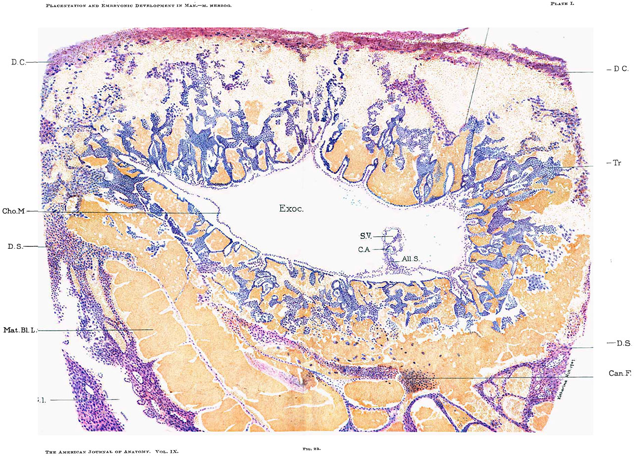
Fig. 23.
Colored plate drawn by Miss Katharina Hill, artist of the Department of Anatomy of the University of Chicago, from an enlargement of fig. 25, photomicrograph from section 153.
The embryo shown in this plate is drawn after section No. 155, because in it the yolk sac, embryonic shield proper, allantois and allantoic duct are best seen.
- All. S. — Allantoic stalk.
- C. A. — Cavity of amnion.
- Can. F. — F1ne fibrin threads and leucocytes (early canalized fibrin).
- Cho. M. — Chorion mesoderni.
- D. C. — Decidua capsularis.
- Exoc. — Exocoelom.
- Gl. — Gland space (epithelium partly preserved).
- Mat. Bl. S. — Large cystic gland spaces filled with blood.
- S. V. — Yolk sac.
- Syn. — Syncytium.
- Tr. — Trophoblast.
| Historic Disclaimer - information about historic embryology pages |
|---|
 |
Reference
Herzog MA. A contribution to our knowledge of the earliest known stages of placentation and embryonic development in man. (1909) Amer. J Anat., 9(3): 361-400.
Cite this page: Hill, M.A. (2024, April 28) Embryology Herzog1909 plate01.jpg. Retrieved from https://embryology.med.unsw.edu.au/embryology/index.php/File:Herzog1909_plate01.jpg
- © Dr Mark Hill 2024, UNSW Embryology ISBN: 978 0 7334 2609 4 - UNSW CRICOS Provider Code No. 00098G
File history
Click on a date/time to view the file as it appeared at that time.
| Date/Time | Thumbnail | Dimensions | User | Comment | |
|---|---|---|---|---|---|
| current | 16:19, 22 November 2016 | | 1,280 × 922 (441 KB) | Z8600021 (talk | contribs) | |
| 16:19, 22 November 2016 |  | 3,752 × 2,751 (1.72 MB) | Z8600021 (talk | contribs) | ==Fig. 23.== Colored plate drawn by Miss Katharina Hill, artist of the Department of Anatomy of the University of Chicago, from an enlargement of fig. 25, photomicrograph from section 153. The embryo shown in this plate is drawn after section No. 155... |
You cannot overwrite this file.
File usage
The following page uses this file: