File:HertigAdams1967 fig01-4.jpg

Original file (1,691 × 2,353 pixels, file size: 390 KB, MIME type: image/jpeg)
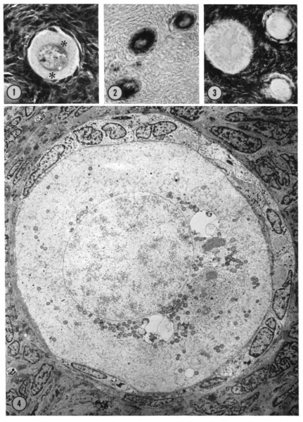
FIGURE 1 Mid—polar section through an oocyte in a primordial follicle. Closely applied to the spherical vesicular nucleus is the crescent—shaped Balbiani’s vitelline body (between asterisks at lower right against nucleus) formed by peripheral dense granules (mitochondria) and a central paler area (cytoeentrum) surrounding a dense body. H 48, H —l— 14}. X 560.
FIGURE 2 Note that the cytoplasm of these three oocytes in primordial follicles is strongly reactive for glucose—6—phosphate dehydrogenase. II 54; paraformaldehyde fixative. x 350.
FIGURE 3 The cortical cells and the periphery of the cells in the Wall of the primitive follicles at right are reactive for adenosine~Inonophosphatase. The pronounced activity at the oocyte—follicle cell junction apparently is located in the numerous projections of the follicle cells present in this intereellula.r space. The activity of the enzyme is not maintained at the periphery of differentiating follicular cells of the multilaminous primary follicle at left. H 53; paraformaldehyde fixative. X 350.
FIGURE 4 A low—power survey micrograph through the mid-polar axis of an oocyte in a primordial follicle. Note that most of the organelles are concentrated in one pole of the oocyte; compare with Fig. 9.5. II 44-1; Karnovsky fixative; uranyl and lead stain. X 2700.
| Abbreviations | ||||
|---|---|---|---|---|
| AL, annulate lamellae
C, Cortex CA, compound aggregates |
CF, coarse fiber
CH, cell chromatin F, follicle cell |
G, Golgi complex
H, heterochromatin I, interchromatin granules |
MT, microtubules
N, nucleus NU, nucleolus |
O, oocyte
P, projection VA, vesicular aggregates |
| Note: In the legends the II number which identifies each patient is followed by another number identifying the particular oocyte represented. | ||||
Reference
Hertig AT. and Adams EC. Studies on the human oocyte and its follicle. I. Ultrastructural and histochemical observations on the primordial follicle stage. (1967) J Cell Biol. 34(2):647-75. PMID 4292010
Copyright
Rockefeller University Press - Copyright Policy This article is distributed under the terms of an Attribution–Noncommercial–Share Alike–No Mirror Sites license for the first six months after the publication date (see http://www.jcb.org/misc/terms.shtml). After six months it is available under a Creative Commons License (Attribution–Noncommercial–Share Alike 4.0 Unported license, as described at https://creativecommons.org/licenses/by-nc-sa/4.0/ ). (More? Help:Copyright Tutorial)
Cite this page: Hill, M.A. (2024, April 27) Embryology HertigAdams1967 fig01-4.jpg. Retrieved from https://embryology.med.unsw.edu.au/embryology/index.php/File:HertigAdams1967_fig01-4.jpg
- © Dr Mark Hill 2024, UNSW Embryology ISBN: 978 0 7334 2609 4 - UNSW CRICOS Provider Code No. 00098G
File history
Click on a date/time to view the file as it appeared at that time.
| Date/Time | Thumbnail | Dimensions | User | Comment | |
|---|---|---|---|---|---|
| current | 17:55, 1 May 2018 |  | 1,691 × 2,353 (390 KB) | Z8600021 (talk | contribs) | ===Reference=== {{Ref-HertigAdams1967}} {{JCB}} {{Footer}} |
You cannot overwrite this file.
File usage
The following page uses this file: