File:Hertig1956 plate17.jpg

Original file (1,482 × 2,228 pixels, file size: 461 KB, MIME type: image/jpeg)
Plate 17. 12-day Previllous Ovum
The implantation site of a 12-day previllous ovum, normal except for a shallow type of implantation.
87 Surface view of the intact implantation site showing the excessive hemorrhage above the ovum, best appreciated in the profile view shown in figure 88. Carnegie 8000, Sequence 2. X 5.
88 Profile view of the intact implantation site to show that the excessive hemorrhage completely conceals the shallowly implanted ovum seen in figures 89 and 90. Carnegie 8000, Sequence 3. X 5.
89 A low—power View of a section of the ovum to show the excessive hemorrhage from its normally defective decidua capsularis giving the ovum a more polypoicl appearance than its depth of implantation would actually warrant. Note progestational hyperplasia of endometrium normal for this stage of pregnancy. Carnegie 8000, Section 11-3-3. X 20?“,
90 A medium-power view of a section of the ovum in figures 87-89 to show the essential normality of the specimen, aside from its shallow implantation. It is doubtful whether this pregnancy would have resulted in abortion, although such a shallow implantation might possibly result in the formation of a circumvallate placenta. This anomaly of placental development may in turn cause abortion although many pregnancies whose placentas are of circumxrallate type do go on to term. Carnegie 8000, Section 11-3-2. X 100.
- Abnormal Implantation 12 days
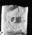
87 Surface view of the intact implantation site
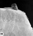
88 Profile view of the intact implantation site
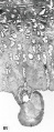
89 A low-power view of a section of the ovum
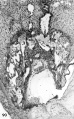
90 A medium-power view of a section of the ovum


- Figure Links: 1 | 2 | 3 | 4 | 5 | 6 | 7 | 8 | 9-10 | 11-12 | 13-14 | 15-16 | 17 | 18-19 | 20 | 21-22 | 23 | 24-25 | 26-27 | 28-29 | 30-31 | 32-33 | 34 | 35 | 36 | 37 | 38 | 39 | 40 | 41 | 42 | 43 | 44 | 45 | 46 | 47 | 48 | 40 | 49 | 50 | 51 | 52 | 53 | 54 | 55 | 56 | 57 | 58 | 59 | 60 | 61 | 62 | 63 | 64 | 65 | 66 | 67 | 68 | 69 | 70 | 71 | 72 | 73 | 74 | 75 | 76 | 77 | 78 | 79 | 80 | 81 | 82 | 83 | 84 | 85 | 86 | 87 | 88 | 89 | 90 | plate 1 | plate 2 | plate 3 | plate 4 | plate 5 | plate 6 | plate 7 | plate 8 | plate 9 | plate 10 | plate 11 | plate 12 | plate 13 | plate 14 | plate 15 | plate 16 | plate 17 | table 1 | table 1 image | table 2 image | table 3 image | table 4 | table 4 image | table 5 | table 5 image | All figures | 1956 Hertig | Embryology History - Arthur Hertig | John Rock | Historic Papers
Reference
Hertig AT. Rock J. and Adams EC. A description of 34 human ova within the first 17 days of development. (1956) Amer. J Anat., 98:435-493.
Cite this page: Hill, M.A. (2024, April 27) Embryology Hertig1956 plate17.jpg. Retrieved from https://embryology.med.unsw.edu.au/embryology/index.php/File:Hertig1956_plate17.jpg
- © Dr Mark Hill 2024, UNSW Embryology ISBN: 978 0 7334 2609 4 - UNSW CRICOS Provider Code No. 00098G
File history
Click on a date/time to view the file as it appeared at that time.
| Date/Time | Thumbnail | Dimensions | User | Comment | |
|---|---|---|---|---|---|
| current | 23:06, 22 February 2017 | | 1,482 × 2,228 (461 KB) | Z8600021 (talk | contribs) | |
| 23:06, 22 February 2017 |  | 1,561 × 2,471 (554 KB) | Z8600021 (talk | contribs) | {{Hertig1956 figures}} {{Ref-Hertig1956}} |
You cannot overwrite this file.
File usage
The following page uses this file: