File:Hertig1956 plate12.jpg

Original file (1,280 × 1,894 pixels, file size: 293 KB, MIME type: image/jpeg)
Plate 12
Four abnormal ova of the pre-implantation stage. They all appear to be 3 to 5 days of age on the basis of coital data, endometrial findings and ovular development. They have, in common, the abnormality of nulltinucleated blastomeres. In addition some show variable degrees of cellular degeneration or necrobiosis and it appears doubtful that they would have retained the ability to implant. even though surgery had not intervened.
59 An intact abnormal 5-cell morula showing variation in the size and shape of the blastomeres each of which contains a number of small rounded nuclear fragments. Note intact zona closely applied to the blastomeres, an artifact of fixation. Carnegie 8630, Sequence 7. X 500.
60 A section of the ovum shown in figure 59 to sho\v the variation of nuclear form. The blastomere at the left contains numerous small rounded nuclei or nuclear fragments. The middle blastomere contains a large indistinct nucleolus and the one on the right contains three pairs of chromatin masses that strongly suggest, a disintegrating mitotic figure of triaster type. Note two polar bodies below and artifactitiously ruptured zona. Carnegie 8630, Section 9. X 500.
61 An intact abnormal 8-cell morula to show its flattened shape. When viewed from above as an intact object it was disc-like with some blastomeres obviously multinueleated and others indistinct and foamy. Carnegie 8450,, Sequence ll. X 500.
62 A section through the greatest diameter of the ovum shown in figure 61. Note the two nearly normal blastomeres on the right contrasted with the two paler ones whose nuclei are undergoing degeneration. Other blastomeres not shown in this section were. multinucleate and/or indistinct with nuclear masses or dense nuclear fragments. Carnegie 8450, Section 8. X 500.
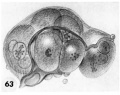
63 A drawing of a glass-plate reconstruction of a 9-cell abnormal morula. Note artifactitious rupture of zona, unexplained cellular fragments and nuclear detail in the various blastomeres. Carnegie 8190. X 500.
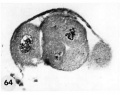
64 A section of the ovum shown in figure 63. Note variation in size and shape of the blastomeres (many in other sections were multinueleated), the cleft. which may indicate early formation of a segmentation cavity and the mitotic figure. Carnegie 8190, Section 5. X 500.
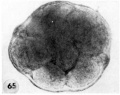
65 An intact abnormal 11- to 12-cell morula showing the intact zona, the position of the two polar bodies and the size, shape and arrangement of the blastomeres.Carnegie 8452 Sequence 3. X 500.
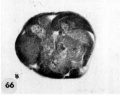
66 A section through the greatest diameter of the ovum shown in figure 65. Note intact zona, the polar body and the variation in size, shape and staining reaction of the blastomeres. The pale, larger cells may be of potential embryonic nature, whereas the darker irregular multinucleated ones may be of future trophoblastic type. Carnegie 8452, Section 8. X 500.
- Abnormal 3 to 5 days
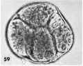
59 abnormal 5-cell morula
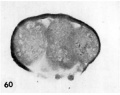
60 section abnormal 5-cell morula
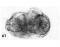
61 abnormal 8-cell morula
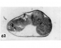
62 section abnormal 8-cell morula
63 abnormal 9-cell morula
64 section abnormal 9-cell morula
65 abnormal 11- to 12-cell morula
66 section abnormal 11- to 12-cell morula


- Figure Links: 1 | 2 | 3 | 4 | 5 | 6 | 7 | 8 | 9-10 | 11-12 | 13-14 | 15-16 | 17 | 18-19 | 20 | 21-22 | 23 | 24-25 | 26-27 | 28-29 | 30-31 | 32-33 | 34 | 35 | 36 | 37 | 38 | 39 | 40 | 41 | 42 | 43 | 44 | 45 | 46 | 47 | 48 | 40 | 49 | 50 | 51 | 52 | 53 | 54 | 55 | 56 | 57 | 58 | 59 | 60 | 61 | 62 | 63 | 64 | 65 | 66 | 67 | 68 | 69 | 70 | 71 | 72 | 73 | 74 | 75 | 76 | 77 | 78 | 79 | 80 | 81 | 82 | 83 | 84 | 85 | 86 | 87 | 88 | 89 | 90 | plate 1 | plate 2 | plate 3 | plate 4 | plate 5 | plate 6 | plate 7 | plate 8 | plate 9 | plate 10 | plate 11 | plate 12 | plate 13 | plate 14 | plate 15 | plate 16 | plate 17 | table 1 | table 1 image | table 2 image | table 3 image | table 4 | table 4 image | table 5 | table 5 image | All figures | 1956 Hertig | Embryology History - Arthur Hertig | John Rock | Historic Papers
Reference
Hertig AT. Rock J. and Adams EC. A description of 34 human ova within the first 17 days of development. (1956) Amer. J Anat., 98:435-493.
Cite this page: Hill, M.A. (2024, April 27) Embryology Hertig1956 plate12.jpg. Retrieved from https://embryology.med.unsw.edu.au/embryology/index.php/File:Hertig1956_plate12.jpg
- © Dr Mark Hill 2024, UNSW Embryology ISBN: 978 0 7334 2609 4 - UNSW CRICOS Provider Code No. 00098G
File history
Click on a date/time to view the file as it appeared at that time.
| Date/Time | Thumbnail | Dimensions | User | Comment | |
|---|---|---|---|---|---|
| current | 14:41, 24 February 2017 | | 1,280 × 1,894 (293 KB) | Z8600021 (talk | contribs) | |
| 14:39, 23 February 2017 | 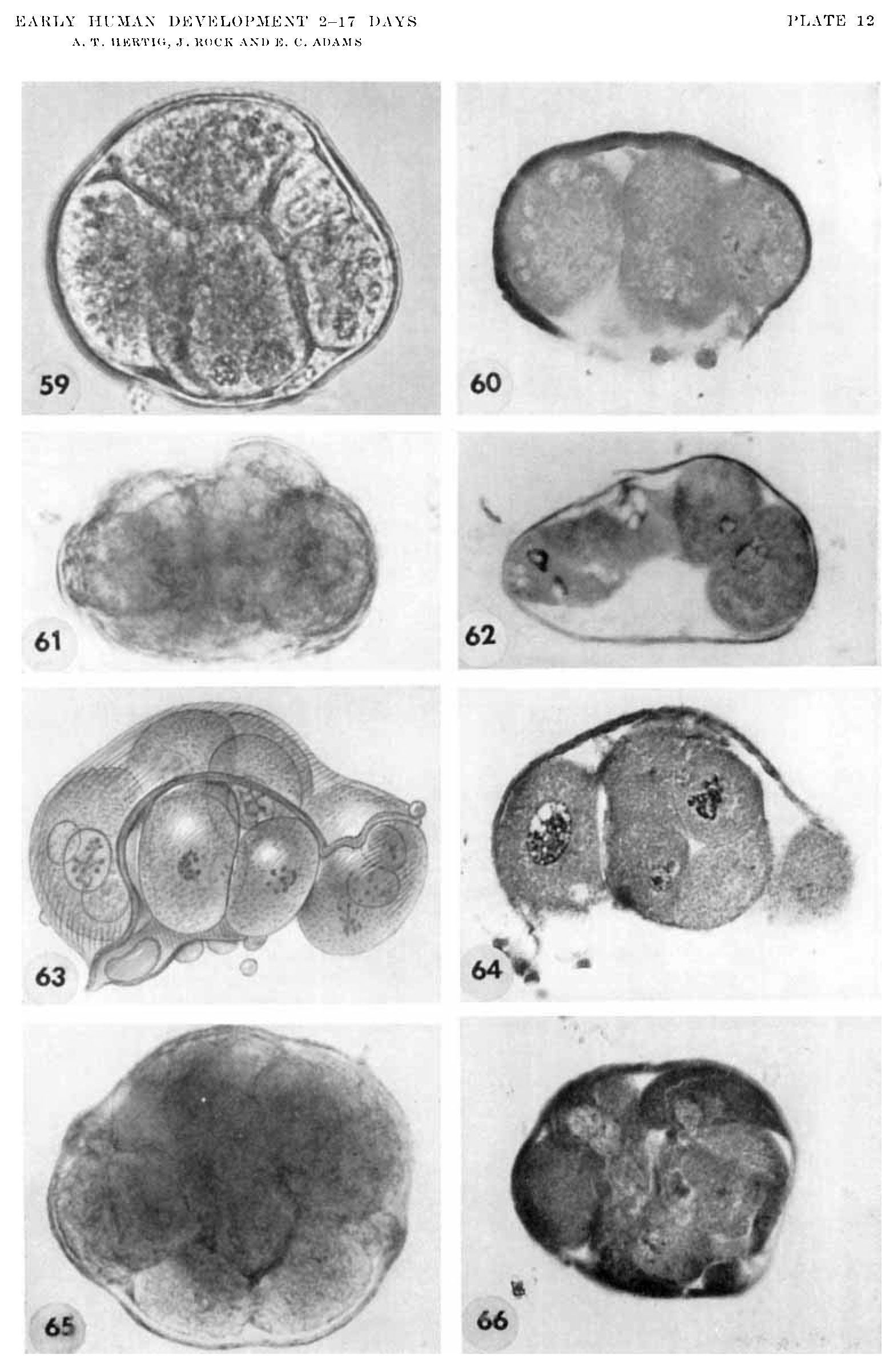 | 1,564 × 2,404 (389 KB) | Z8600021 (talk | contribs) |
You cannot overwrite this file.
File usage
The following page uses this file: