File:Hertig1956 plate11.jpg

Original file (1,280 × 1,909 pixels, file size: 458 KB, MIME type: image/jpeg)
Plate 11
Drawings made from transparent three-dimensional plastic reconstructions of ova of 7, 8, 9, 11 and 12 days of age.
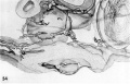
54 Reconstruction of half a 7-day ovum, which presents an uneven surface of its solid trophohlast (stippled) to the adjacent endometrium. The trophoblast has already surrounded a. capillary sinusoid (left). The cut surface of the hilaminar germ disc is evident (endoderm cellular, ectoderm solid). Note primitive amniogenic cells above tiny amniotic cavity. The maternal sinusoidal network is merely the arteriovenous capillary network of the endometrium that has begun to undergo hypertrophy and dilation. See figures I1 and 12. Carnegie 8020. X 260.
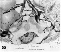
55 Reconstruction of half an 8-day ovum viewed from the level of the. greatest diameter of the embryo. The top section of the reconstruction coincides with the photomicrograph of this ovum in figure 13. The trophoblast (stippled) is still solid and is encircling the tips of maternal siuusoids which, although prominent, actually contain very little blood. The large sinusoid toward the right is interpreted as venous in type, whereas the coiled smaller one to the left is a spiral arteriole. The existing capillary anastomoses between this endometrial arteriole and vcnule are the structures that actually grow and dilate under the stimulus of the immediately adjacent trophoblast. Note amniotic cavity but lack of amnioblasts. dorsally concave hilaminar germ disc and mesoblasts within the chorionic cavity. See figures 13 and 14. Carnegie 8155. X 200.
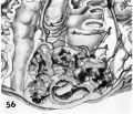
56 Reconstruction of half a 9-day ovum and surrounding endometrium, viewed from the level of the section illustrated in figure 31. Note bilamiuar germ disc and amnion still attached at one point to the overlying trophoblast. Although the trophohlastic lacunae appear discontinuous on the cut surface they actually are a continuous laeunar network in the depths of the trophoblast. Note several points at which the surrounding endometrial sinasoids communicate with this laeunar network. The latter is still ischemie presumably due to spasm of the arteriolar blood supply. See figures 21-23. Carnegie 8004. X 100.
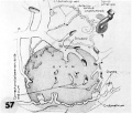
57 A non-schematic line drawing of an 11-day ovum to illustrate the uterolacanar circulation at this developmental stage. In contrast to the cross-sectional views of the other reconstructions on this plate, this is a view of the external aspect of the ovum. Note the vein and spiral arteriole with arteriovenosus capillary anastomoses. The growth and dilatation of the large sinusoid continuous with and derived from such an anastomosis are presumably due to the presence of the adjacent trophoblast. Note that the glands are represented merely as stumps to allow uninterrupted view of the utero-laeunar circulation. See figures 24, :35 and 34. Carnegie 7699. X 50.
58 Reconstruction of half a 12-day ovum to illustrate the details of the embryo and chorionic cavity. The top surface of the reconstruction corresponds to the section in figure 29. Note intercommunicating nature of laeunar space which is confluent with capillaries of the large arteriovenous plexus in the surrounding endometrium. The filmy trabeculae of the cxtraembryonic mesoblast. support the exocoelomic membrane (thus accounting for the shape of its enclosed cavity), germ disc and cleft-like amniotic cavity. See figures 28, 29 and 36. Carnegie 7700. X 80.
- 7, 8, 9, 11 and 12 days
54 Reconstruction half a 7-day ovum
55 Reconstruction half an 8-day ovum
56 Reconstruction half a 9-day ovum
57 A non-schematic line drawing 11-day ovum
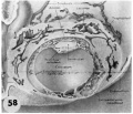
58 Reconstruction half a 12-day ovum


- Figure Links: 1 | 2 | 3 | 4 | 5 | 6 | 7 | 8 | 9-10 | 11-12 | 13-14 | 15-16 | 17 | 18-19 | 20 | 21-22 | 23 | 24-25 | 26-27 | 28-29 | 30-31 | 32-33 | 34 | 35 | 36 | 37 | 38 | 39 | 40 | 41 | 42 | 43 | 44 | 45 | 46 | 47 | 48 | 40 | 49 | 50 | 51 | 52 | 53 | 54 | 55 | 56 | 57 | 58 | 59 | 60 | 61 | 62 | 63 | 64 | 65 | 66 | 67 | 68 | 69 | 70 | 71 | 72 | 73 | 74 | 75 | 76 | 77 | 78 | 79 | 80 | 81 | 82 | 83 | 84 | 85 | 86 | 87 | 88 | 89 | 90 | plate 1 | plate 2 | plate 3 | plate 4 | plate 5 | plate 6 | plate 7 | plate 8 | plate 9 | plate 10 | plate 11 | plate 12 | plate 13 | plate 14 | plate 15 | plate 16 | plate 17 | table 1 | table 1 image | table 2 image | table 3 image | table 4 | table 4 image | table 5 | table 5 image | All figures | 1956 Hertig | Embryology History - Arthur Hertig | John Rock | Historic Papers
Reference
Hertig AT. Rock J. and Adams EC. A description of 34 human ova within the first 17 days of development. (1956) Amer. J Anat., 98:435-493.
Cite this page: Hill, M.A. (2024, April 27) Embryology Hertig1956 plate11.jpg. Retrieved from https://embryology.med.unsw.edu.au/embryology/index.php/File:Hertig1956_plate11.jpg
- © Dr Mark Hill 2024, UNSW Embryology ISBN: 978 0 7334 2609 4 - UNSW CRICOS Provider Code No. 00098G
File history
Click on a date/time to view the file as it appeared at that time.
| Date/Time | Thumbnail | Dimensions | User | Comment | |
|---|---|---|---|---|---|
| current | 22:42, 23 February 2017 | | 1,280 × 1,909 (458 KB) | Z8600021 (talk | contribs) | |
| 14:39, 23 February 2017 | 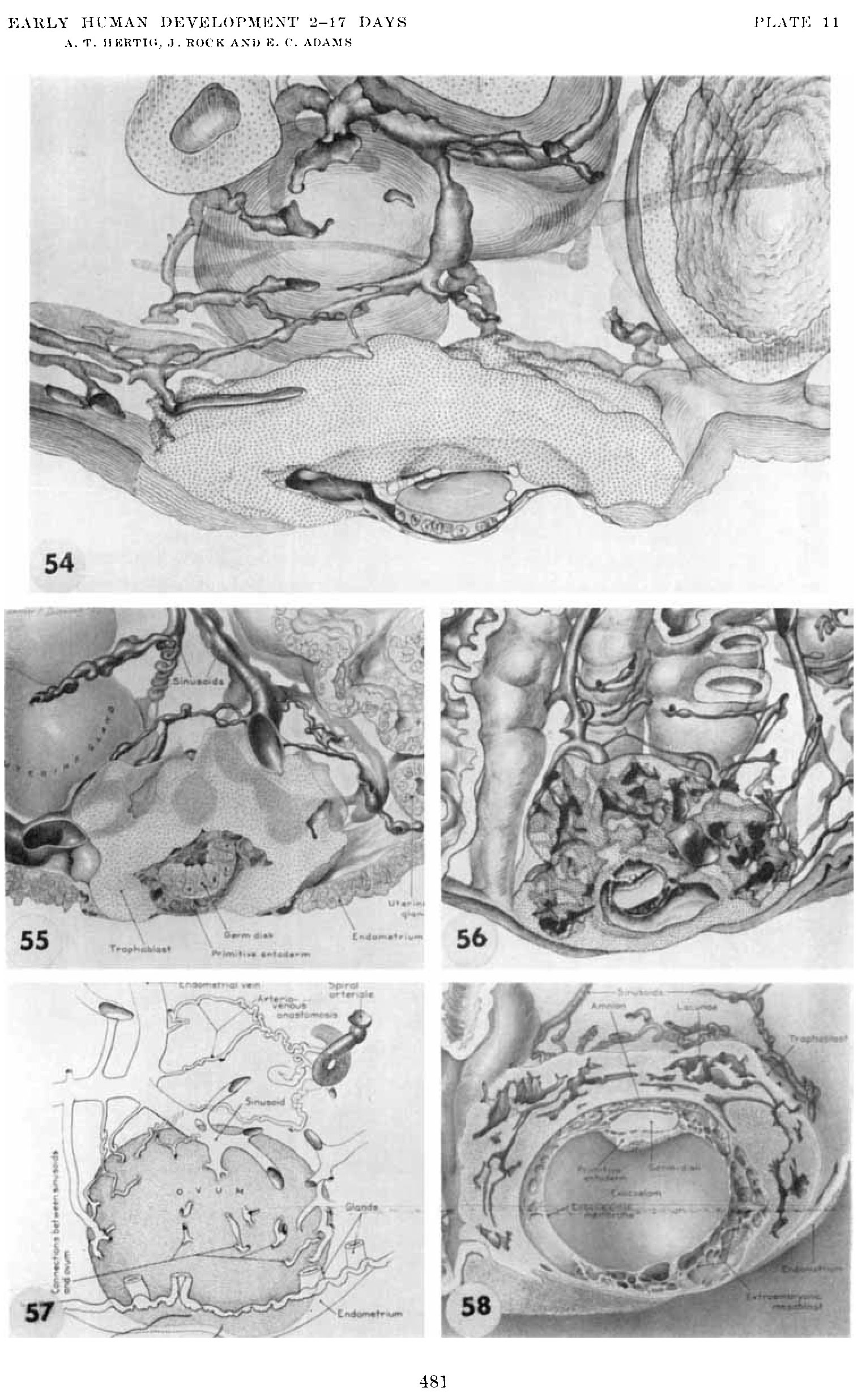 | 1,529 × 2,471 (650 KB) | Z8600021 (talk | contribs) |
You cannot overwrite this file.
File usage
The following page uses this file: