File:Hertig1956 plate10.jpg

Original file (1,000 × 1,912 pixels, file size: 461 KB, MIME type: image/jpeg)
Plate 10
Low-power views of sections from the two villous ova illustrated on plate 9.
52 A 16-day ovum showing branched villi on the entire external surface‘ of the chorion. Note that the cytotrophoblast at the peripheral tips of the villi is beginning to coalesce to form the plat-o11t:1l floor which is peiietrated by nmternal blood vessels of the utero-placental circulation. Note large sinusoid at upper right. The decidual reaction around the entire surface of ovum is prominent, thus foreshadowing the formtion of the decidual basalis and capsularis. Carnegie 7802, Section 4-4~3—5. X 30.
53 A 17-day ovum showing the further development of the branching villi with their anchorage to the eytotrophoblust of the placental floor. Note expanded intervillous space connecting with numerous prominent thin-walled maternal sinusoids thus constituting the utero-placental circulation. This is the beginning of :1 more active flow of innternzil blood through the placenta although small but increasing amounts of such blood have been present since the 10th (lay. Note also the decidua around the ovum with many darkly staining tlettielied syncytiotrophoblastic giant cells. Carnegie 8602, Section 25—3—2. X 30.
- Streeter Horizon VII
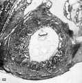
52 A 16-day ovum
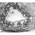
53 A 17-day ovum


- Figure Links: 1 | 2 | 3 | 4 | 5 | 6 | 7 | 8 | 9-10 | 11-12 | 13-14 | 15-16 | 17 | 18-19 | 20 | 21-22 | 23 | 24-25 | 26-27 | 28-29 | 30-31 | 32-33 | 34 | 35 | 36 | 37 | 38 | 39 | 40 | 41 | 42 | 43 | 44 | 45 | 46 | 47 | 48 | 40 | 49 | 50 | 51 | 52 | 53 | 54 | 55 | 56 | 57 | 58 | 59 | 60 | 61 | 62 | 63 | 64 | 65 | 66 | 67 | 68 | 69 | 70 | 71 | 72 | 73 | 74 | 75 | 76 | 77 | 78 | 79 | 80 | 81 | 82 | 83 | 84 | 85 | 86 | 87 | 88 | 89 | 90 | plate 1 | plate 2 | plate 3 | plate 4 | plate 5 | plate 6 | plate 7 | plate 8 | plate 9 | plate 10 | plate 11 | plate 12 | plate 13 | plate 14 | plate 15 | plate 16 | plate 17 | table 1 | table 1 image | table 2 image | table 3 image | table 4 | table 4 image | table 5 | table 5 image | All figures | 1956 Hertig | Embryology History - Arthur Hertig | John Rock | Historic Papers
Reference
Hertig AT. Rock J. and Adams EC. A description of 34 human ova within the first 17 days of development. (1956) Amer. J Anat., 98:435-493.
Cite this page: Hill, M.A. (2024, April 27) Embryology Hertig1956 plate10.jpg. Retrieved from https://embryology.med.unsw.edu.au/embryology/index.php/File:Hertig1956_plate10.jpg
- © Dr Mark Hill 2024, UNSW Embryology ISBN: 978 0 7334 2609 4 - UNSW CRICOS Provider Code No. 00098G
File history
Click on a date/time to view the file as it appeared at that time.
| Date/Time | Thumbnail | Dimensions | User | Comment | |
|---|---|---|---|---|---|
| current | 22:29, 23 February 2017 | | 1,000 × 1,912 (461 KB) | Z8600021 (talk | contribs) | |
| 14:38, 23 February 2017 |  | 1,434 × 2,387 (525 KB) | Z8600021 (talk | contribs) |
You cannot overwrite this file.
File usage
The following 2 pages use this file: