File:Hertig1956 plate08.jpg

Original file (1,280 × 1,987 pixels, file size: 497 KB, MIME type: image/jpeg)
Plate 8
Sections at low-power magnification of the three 13-day ova of Horizon VI shown on plate 7. They are characterized by primitive unbranched villi and a distinct yolk-sac.
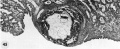
45 A 13-day ovum with a yolk-sac and beginning villi. Note undulating margin of chorionic cavity as the extraembryonic mesoblast forms shallow, broad cores of the villi. Also note yolk-sac formzition with remnants of the disrupted transitory exocoelomic membrane lying in the chorionic cavity ventral to the embryo. Note further the early decidual reaction imniedititely around the ovum. Carnegie Embryo 8672, Section 11-3-2. X 35.
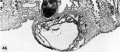
46 A 13-day ovum with yolk-sac and beginning villi. Note decidual reaction and diluted sinusoids. Also note blood-filled gland lumen at the base of the ovum. This is a frequent finding at this stage of development and is due to leakage of blood from the lncunur spaces into the gland lumen following erosion of the gland epithelium by the invading trophohlast. It is often mistaken for a thrombosed blood vessel. Carnegie Embryo 8360, Section 27-4-1. X 35.
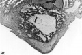
47 A 13-day ovum with unbranched villi and yolk~s:ie. Note remnant of exocoelomic cavity at abembryonic pole of chorionic cavity, many villi and evidence of more active circulation within the lacunar (now properly termed. intervillous) space. Note also the distended and blood-filled gland at the base of the ovum and the coagulated plasma (“Schlussconguluni” of Peters) at the embryonic pole. This maternal blood and plasma, leaking out from the intervillous space, is continuous with the gross blood clot seen in figure 43. Numerous detached darkly staining syncytial giant cells are scattered among the decidual cells immediately surrounding the ovum. Carnegie Embryo 7801, Section 12-1-1. X 35.
- Streeter Horizon VI
45 13-day ovum with a yolk-sac
46 13-day ovum
47 13-day ovum


- Figure Links: 1 | 2 | 3 | 4 | 5 | 6 | 7 | 8 | 9-10 | 11-12 | 13-14 | 15-16 | 17 | 18-19 | 20 | 21-22 | 23 | 24-25 | 26-27 | 28-29 | 30-31 | 32-33 | 34 | 35 | 36 | 37 | 38 | 39 | 40 | 41 | 42 | 43 | 44 | 45 | 46 | 47 | 48 | 40 | 49 | 50 | 51 | 52 | 53 | 54 | 55 | 56 | 57 | 58 | 59 | 60 | 61 | 62 | 63 | 64 | 65 | 66 | 67 | 68 | 69 | 70 | 71 | 72 | 73 | 74 | 75 | 76 | 77 | 78 | 79 | 80 | 81 | 82 | 83 | 84 | 85 | 86 | 87 | 88 | 89 | 90 | plate 1 | plate 2 | plate 3 | plate 4 | plate 5 | plate 6 | plate 7 | plate 8 | plate 9 | plate 10 | plate 11 | plate 12 | plate 13 | plate 14 | plate 15 | plate 16 | plate 17 | table 1 | table 1 image | table 2 image | table 3 image | table 4 | table 4 image | table 5 | table 5 image | All figures | 1956 Hertig | Embryology History - Arthur Hertig | John Rock | Historic Papers
Reference
Hertig AT. Rock J. and Adams EC. A description of 34 human ova within the first 17 days of development. (1956) Amer. J Anat., 98:435-493.
Cite this page: Hill, M.A. (2024, April 27) Embryology Hertig1956 plate08.jpg. Retrieved from https://embryology.med.unsw.edu.au/embryology/index.php/File:Hertig1956_plate08.jpg
- © Dr Mark Hill 2024, UNSW Embryology ISBN: 978 0 7334 2609 4 - UNSW CRICOS Provider Code No. 00098G
File history
Click on a date/time to view the file as it appeared at that time.
| Date/Time | Thumbnail | Dimensions | User | Comment | |
|---|---|---|---|---|---|
| current | 21:58, 23 February 2017 | | 1,280 × 1,987 (497 KB) | Z8600021 (talk | contribs) | |
| 14:38, 23 February 2017 | 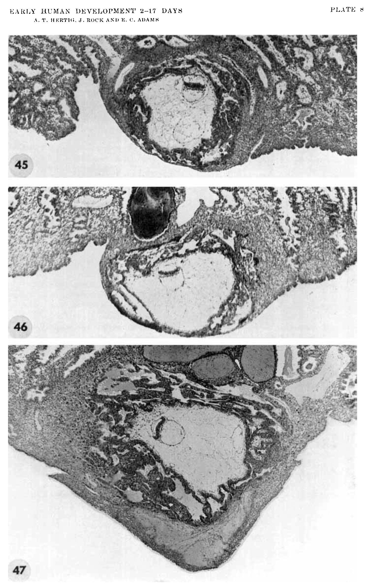 | 1,485 × 2,389 (635 KB) | Z8600021 (talk | contribs) |
You cannot overwrite this file.
File usage
The following page uses this file: