File:Hertig1956 plate07.jpg

Original file (1,481 × 2,215 pixels, file size: 556 KB, MIME type: image/jpeg)
Plate 7
Three ova of Horizon VI about 13 days of age cliaraeterized by primitive villi and a distinct yolk-sac. Low power views of mid sections of each of these ova are shown on plate 8.
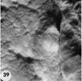
39 Surface view of an intact 13-day ovum found on the posterior uterine wall adjacent to the left lateral sulcns. Note that the endometrial surface has an undulating wrinkled appearance and that the gland mouths are prominent only around the ovum. 8672, Sequence 2. X 8.
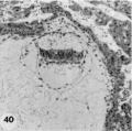
40 Medium-power view of one of the middle sections of the ovum seen in figure 39 to show detail of the embryo and adjacent trophoblast-. Note small double-layer yolk-sac. No villi are seen in this photograph but they are evident at tho ahembryonic pole as shown in figure 45. Note increase in size of amniotic cavity. Its continuity with the trophoblast may be evidence of continuing mnniogenesis from this tissue. Vacuoles are not apparent in the ectoderm but are present in the endoderm. Carnegie Embryo 8672, Section 11-3-2. X 100.
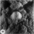
41 Surface view of an intact implantation site of about 13 days. Note smooth dome-shaped elevation of ovum and the marked undnlation of cndometrial surface with fine and coarse wrinkling. '1‘he stellate crevices represent closed gland mouths. Carnegie Embryo 8360, Sequence 3. X 8.
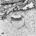
42 A medium-power view of one of the middle sections of the ovum seen in figure 41. Note that the yolk-sac is single-layered, the ectoderm non-vacuolated (a usual feature from now on), and the extrnembryonie mesoblast is prominent in the area lateral and dorsal to the amnion. Carnegie Embryo 8360, Section 27-4-2. X 100.
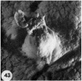
43 Surface view of a 13-day ovum. Note large tab of coagulated maternal blood (a variable but normal feature at this stage) which has escaped from the underlying trophoblastic lacunae through the endomctrial defect. The undulnted and wrinkled surface endometrium is now beginning to resemble decidua. Carnegie Embryo 7801, Sequence 2. X 8.
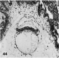
44 A medium-power view of a mid~cross section of the ovum shown in figure 43. Note that the eetoderm laterally has a dorsally curved margin and that the yolk-sac is double-layered. The stulJb_v, unbranched primitive villi have formed about the entire periphery of the chorion and contain extraembryonic mesoblast or primitive fibroblastic tissue. The peripheral tips of these villi will extend outwardly to the endometrium and, as they coalesce at the placental floor, form the anchorage for the developing ovum. Axial differentiation is just appearing in this embryo. The primordium of the primitive streak and a few cells of body mcsoderm are found in sections more cnudad than the one illustrated. Carnegie Embryo 7801, Section 12-1-1. X 100.
- Streeter Horizon VI
39 surface view 13-day ovum
40 middle section 13-day ovum
41 surface view 13-day ovum
42 middle section 13-day ovum
43 surface view 13-day ovum
44 mid~cross section 13-day ovum


- Figure Links: 1 | 2 | 3 | 4 | 5 | 6 | 7 | 8 | 9-10 | 11-12 | 13-14 | 15-16 | 17 | 18-19 | 20 | 21-22 | 23 | 24-25 | 26-27 | 28-29 | 30-31 | 32-33 | 34 | 35 | 36 | 37 | 38 | 39 | 40 | 41 | 42 | 43 | 44 | 45 | 46 | 47 | 48 | 40 | 49 | 50 | 51 | 52 | 53 | 54 | 55 | 56 | 57 | 58 | 59 | 60 | 61 | 62 | 63 | 64 | 65 | 66 | 67 | 68 | 69 | 70 | 71 | 72 | 73 | 74 | 75 | 76 | 77 | 78 | 79 | 80 | 81 | 82 | 83 | 84 | 85 | 86 | 87 | 88 | 89 | 90 | plate 1 | plate 2 | plate 3 | plate 4 | plate 5 | plate 6 | plate 7 | plate 8 | plate 9 | plate 10 | plate 11 | plate 12 | plate 13 | plate 14 | plate 15 | plate 16 | plate 17 | table 1 | table 1 image | table 2 image | table 3 image | table 4 | table 4 image | table 5 | table 5 image | All figures | 1956 Hertig | Embryology History - Arthur Hertig | John Rock | Historic Papers
Reference
Hertig AT. Rock J. and Adams EC. A description of 34 human ova within the first 17 days of development. (1956) Amer. J Anat., 98:435-493.
Cite this page: Hill, M.A. (2024, April 27) Embryology Hertig1956 plate07.jpg. Retrieved from https://embryology.med.unsw.edu.au/embryology/index.php/File:Hertig1956_plate07.jpg
- © Dr Mark Hill 2024, UNSW Embryology ISBN: 978 0 7334 2609 4 - UNSW CRICOS Provider Code No. 00098G
File history
Click on a date/time to view the file as it appeared at that time.
| Date/Time | Thumbnail | Dimensions | User | Comment | |
|---|---|---|---|---|---|
| current | 21:29, 23 February 2017 | | 1,481 × 2,215 (556 KB) | Z8600021 (talk | contribs) | |
| 14:26, 23 February 2017 |  | 1,575 × 2,380 (518 KB) | Z8600021 (talk | contribs) |
You cannot overwrite this file.
File usage
The following page uses this file: