File:Hertig1956 plate06.jpg
Original file (1,280 × 2,022 pixels, file size: 542 KB, MIME type: image/jpeg)
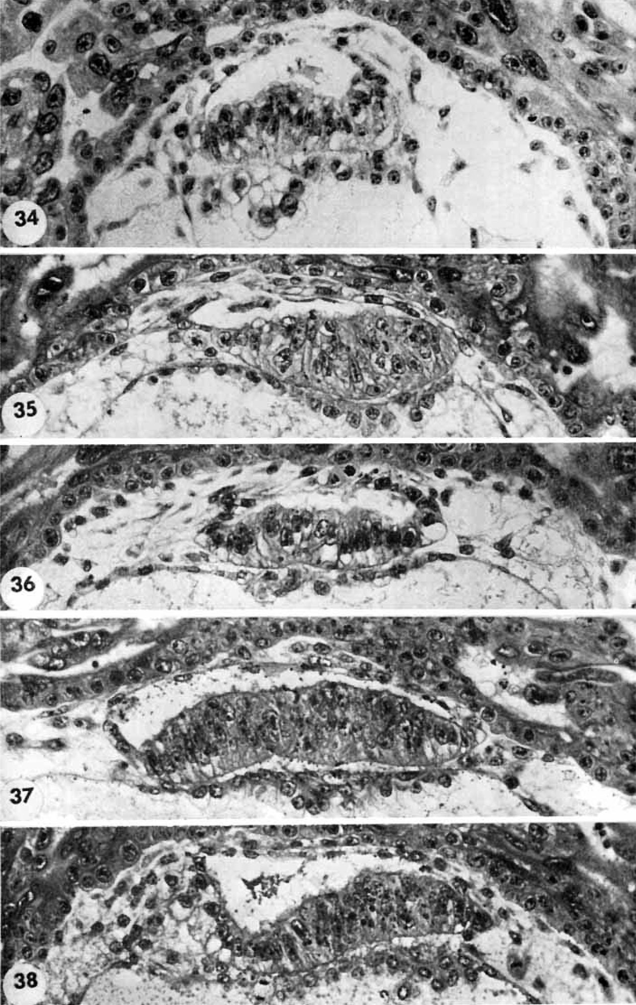
Plate 6
High-power views of the germ discs of the five 11- to 12-day ova of Horizon Vc illustrated previously on plates 4 and 5.
34 The germ disc and anmion of an 11-day ovum. Note the orderly arrangement of cylindrical vacuolated ectodermal cells, beneath which is the irregularly arranged layer of vacuolated and foamy-appearing polyhedral cells of the endoderm. Dorsal to the germ disc is the amnion, delaminated in situ from the overlying cytotrophoblast and attached to the peripheral margin of the ectoderm. Note a. few mesoblasts also of cytotrophoblastic origin external to the amnion and merging with those at the junction of the endodermal plate and the exocoelomic membrane. Carnegie 7699, Section 8-5-3. X 300.
35 The germ disc and amnion of a 12-day ovum. Occasional irregularly distributed vacuoles are seen in the ectoderm. Note the thin layer of polyhedral cells of the endodcrmal plate which joins laterally with the elongated mesohlasts of the exocoelomie membrane. Note the fiat amniotic cavity with amniogenesis from the overlying cytotrophoblasts. Carnegie 7950, Section 11-5-2. X 300.
36 The germ disc and amnion of a 12-day ovum. Note the mitotic figure of the amniotic cell delaminating from the cytotrophoblast. Note also the ventral orientation of the vacuoles in the eetoderm and the less evident dorsal orientation of similar structures in the endoderm. Carnegie 7700, Section 5-7-7. X 300.
37 The germ disc of a 12-day ovum. Note that in this specimen the vacuolated ectodermal cells appear to be located peripherally. Carnegie 8558, Section 10-1-4. X 300.
38 The germ disc of a 12-day ovum. Note that vacuolated ectodermal cells appear to be concentrated toward the left and that the cxtraembryonic. mesoblast is more condensed in this area perhaps indicating the beginning of axis formation in this embryo, the oldest in our Horizon Vc. Carnegie 8330, Section 9-2-5. X 300.
- Streeter Horizon Vc
34 germ disc and anmion 11-day ovum
35 germ disc and amnion 12-day ovum
36 germ disc and amnion 12-day ovum
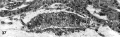
37 germ disc 12-day ovum
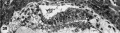
38 germ disc 12-day ovum
- Figure Links: 1 | 2 | 3 | 4 | 5 | 6 | 7 | 8 | 9-10 | 11-12 | 13-14 | 15-16 | 17 | 18-19 | 20 | 21-22 | 23 | 24-25 | 26-27 | 28-29 | 30-31 | 32-33 | 34 | 35 | 36 | 37 | 38 | 39 | 40 | 41 | 42 | 43 | 44 | 45 | 46 | 47 | 48 | 40 | 49 | 50 | 51 | 52 | 53 | 54 | 55 | 56 | 57 | 58 | 59 | 60 | 61 | 62 | 63 | 64 | 65 | 66 | 67 | 68 | 69 | 70 | 71 | 72 | 73 | 74 | 75 | 76 | 77 | 78 | 79 | 80 | 81 | 82 | 83 | 84 | 85 | 86 | 87 | 88 | 89 | 90 | plate 1 | plate 2 | plate 3 | plate 4 | plate 5 | plate 6 | plate 7 | plate 8 | plate 9 | plate 10 | plate 11 | plate 12 | plate 13 | plate 14 | plate 15 | plate 16 | plate 17 | table 1 | table 1 image | table 2 image | table 3 image | table 4 | table 4 image | table 5 | table 5 image | All figures | 1956 Hertig | Embryology History - Arthur Hertig | John Rock | Historic Papers
Reference
Hertig AT. Rock J. and Adams EC. A description of 34 human ova within the first 17 days of development. (1956) Amer. J Anat., 98:435-493.
Cite this page: Hill, M.A. (2024, April 27) Embryology Hertig1956 plate06.jpg. Retrieved from https://embryology.med.unsw.edu.au/embryology/index.php/File:Hertig1956_plate06.jpg
- © Dr Mark Hill 2024, UNSW Embryology ISBN: 978 0 7334 2609 4 - UNSW CRICOS Provider Code No. 00098G
File history
Click on a date/time to view the file as it appeared at that time.
| Date/Time | Thumbnail | Dimensions | User | Comment | |
|---|---|---|---|---|---|
| current | 21:29, 23 February 2017 | | 1,280 × 2,022 (542 KB) | Z8600021 (talk | contribs) | |
| 14:26, 23 February 2017 |  | 1,494 × 2,384 (586 KB) | Z8600021 (talk | contribs) |
You cannot overwrite this file.
File usage
The following page uses this file: