File:Hertig1956 plate05.jpg
Original file (1,280 × 2,002 pixels, file size: 466 KB, MIME type: image/jpeg)
Plate 5
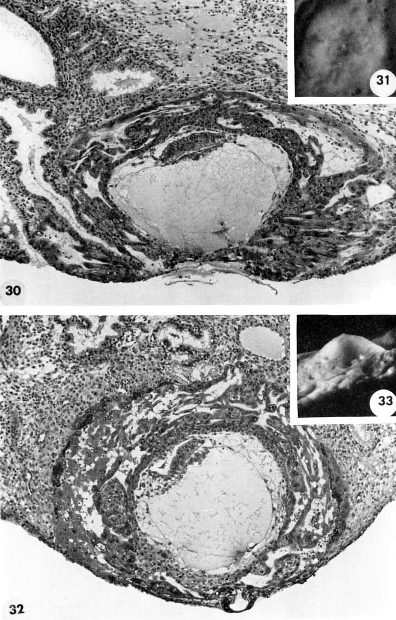
Two normal 12-day ova also in Horizon Vc, characterized by intereommunicating lacunar spaces with evidence of early utero-placental circulation, a distended chorionic cavity, an exocoelomie cavity of maximum size, a bilaminar germ disc and a well-defined amnion and amniotic cavity. Details of their germ discs are shown on plate 6.
30 A medium-power view of a mid-cross section of the ovum shown in figure 31. A coagulum is lying over the uuhealed penetration defect as evidence of seepage of maternal blood and plasma from laennar space into the uterine lumen. Note the persistent edema of the endometrium and the early decidua developing immediately around the ovum. Note primordial villi and also the gland on the left which has been surrounded by syncytimn. A dilated blood vessel is seen at the margin of the ovum at the right. Carnegie 8558, Section 10-1-4. X 100.
31 A surface view of an intact 12-day implantation site to show the endonietrial detect, the source of maternal bleeding into the uterine lumen at. this and slightly later stages. Note the small tabs of coagalum in the ulcer. A hemorrhage, which became detached and hence is not shown, arose from this defect. Carnegie 8558, Sequence 5. X22.
32 A medium-power view of a mid-cross section of the ovum shown in figure 33. Note the early deeidual reaction about the ovum, the primordiuni of a villus on the left, the few blood cells within the lacnnar spaces and the large exocoelomic cavity. Carnegie 8330, Section 9-2-6. X 100.
33 A profile View of a 12-day ovum to show its elevation from the surrounding endometrium. Carnegie 8330, Sequence 3. X 12.
- Streeter Horizon Vc
- Hertig1956 fig31-32.jpg
31-32 12-day ovum
32-33 12-day ovum

- Figure Links: 1 | 2 | 3 | 4 | 5 | 6 | 7 | 8 | 9-10 | 11-12 | 13-14 | 15-16 | 17 | 18-19 | 20 | 21-22 | 23 | 24-25 | 26-27 | 28-29 | 30-31 | 32-33 | 34 | 35 | 36 | 37 | 38 | 39 | 40 | 41 | 42 | 43 | 44 | 45 | 46 | 47 | 48 | 40 | 49 | 50 | 51 | 52 | 53 | 54 | 55 | 56 | 57 | 58 | 59 | 60 | 61 | 62 | 63 | 64 | 65 | 66 | 67 | 68 | 69 | 70 | 71 | 72 | 73 | 74 | 75 | 76 | 77 | 78 | 79 | 80 | 81 | 82 | 83 | 84 | 85 | 86 | 87 | 88 | 89 | 90 | plate 1 | plate 2 | plate 3 | plate 4 | plate 5 | plate 6 | plate 7 | plate 8 | plate 9 | plate 10 | plate 11 | plate 12 | plate 13 | plate 14 | plate 15 | plate 16 | plate 17 | table 1 | table 1 image | table 2 image | table 3 image | table 4 | table 4 image | table 5 | table 5 image | All figures | 1956 Hertig | Embryology History - Arthur Hertig | John Rock | Historic Papers
Reference
Hertig AT. Rock J. and Adams EC. A description of 34 human ova within the first 17 days of development. (1956) Amer. J Anat., 98:435-493.
Cite this page: Hill, M.A. (2024, April 27) Embryology Hertig1956 plate05.jpg. Retrieved from https://embryology.med.unsw.edu.au/embryology/index.php/File:Hertig1956_plate05.jpg
- © Dr Mark Hill 2024, UNSW Embryology ISBN: 978 0 7334 2609 4 - UNSW CRICOS Provider Code No. 00098G
File history
Click on a date/time to view the file as it appeared at that time.
| Date/Time | Thumbnail | Dimensions | User | Comment | |
|---|---|---|---|---|---|
| current | 21:28, 23 February 2017 | | 1,280 × 2,002 (466 KB) | Z8600021 (talk | contribs) | |
| 14:25, 23 February 2017 | 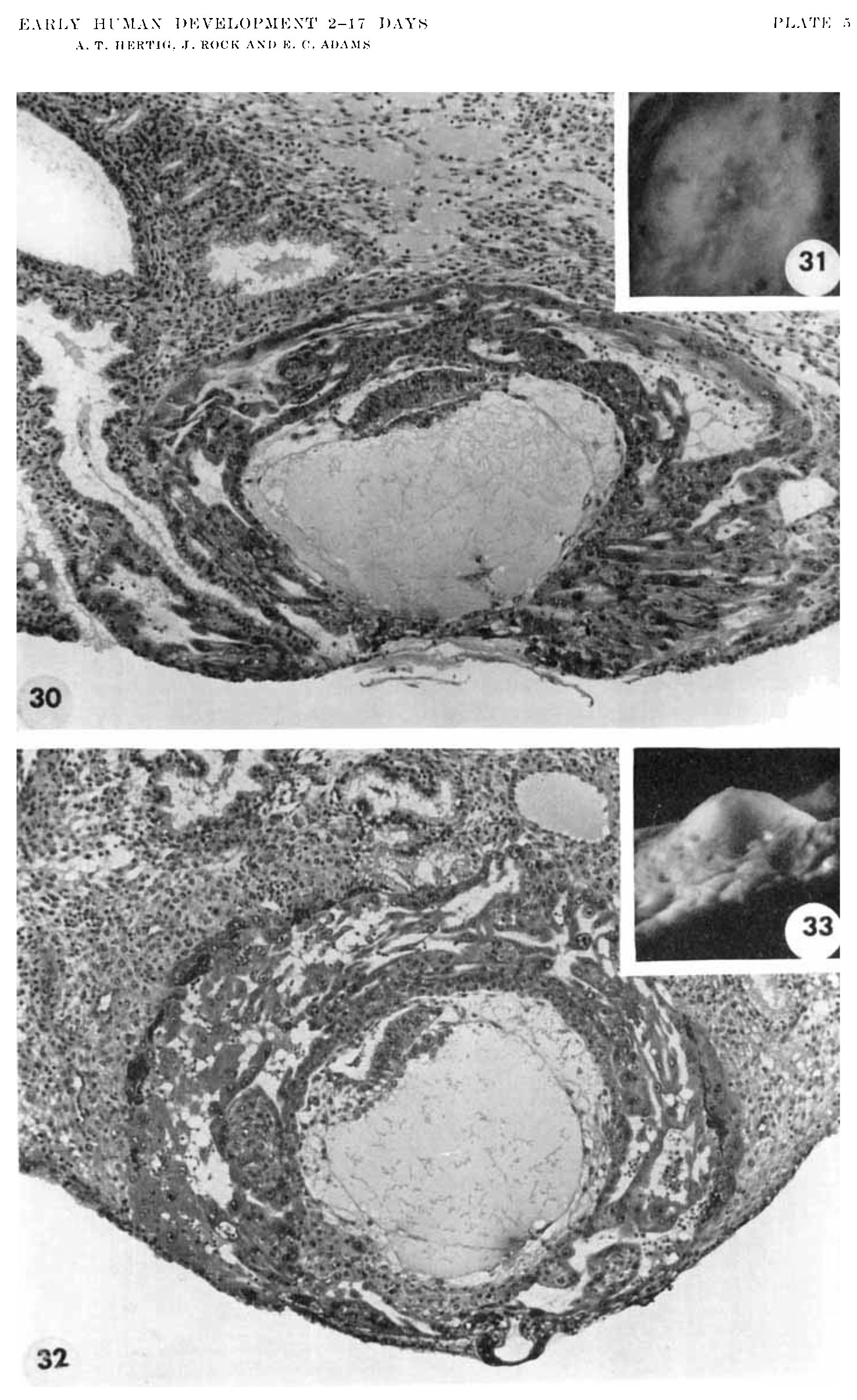 | 1,474 × 2,407 (543 KB) | Z8600021 (talk | contribs) |
You cannot overwrite this file.
File usage
The following page uses this file: