File:Hertig1956 plate04.jpg

Original file (1,280 × 1,904 pixels, file size: 452 KB, MIME type: image/jpeg)
Plate 4
Three normal, late previllous ova 11 to 12 days old, of Horizon Vc. characterized by intereommunieating laeunar spaces filled with maternal plasma containing still increasing numbers of blood cells, a distended chorionic. cavity, an execoelomic cavity of maximum size, a bilaminar germ disc and well~defined amnion and amniotic cavity. Details of the germ discs of these ova are shown on plate 6.
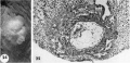
24 Surface view of an intact. 11-day implantation site showing wrinkling of endomctrium between and at the gland mouths. From the endometrial defect created by the ovum arises a. coagulum which communicates with maternal blood within the lacunae. Carnegie 7699, Sequence 8. X 22.
25 A medium-power view of a mid-cross section of the ovum shown in figure 2-}. Note that the endometrial epithelium is proliferating to repair the penetration defect. The cytotrophoblast, immediately surrounding the chorionic cavity, gives rise, by peripheral proliferation and differentiation, to irregular masses which grow into and become invested by syncytiotrophoblast, thus constituting the primordia of chorionic villi. Note syncytium surrounding an endometrial gland and maternal white blood cells in the lacunae. Carnegie 7699, Section 8-5-3. X 100.
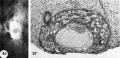
26 Surface view of an intact 12-day-ovum, transilluminated after clearing, showing chorionic cavity as a dark central area with surrounding opaque but vacuolated trophoblast. Note wrinkled endometrial surface with gland months evident as dark pits. Carnegie 7950, Sequence 10. X 22.
27 A medium-power view of a mid-cross section of the ovum shown in figure 26. The edema of the endometrinm, though variable, is characteristic of specimens of this general age. Note the distended exocoelomic cavity, the coalesced lacunar spaces showing evidence of an early utero-placental circulation and the budding of the cytotrophoblast to form primordia. of villi. The penetration defect has not been completely healed. Note the cross-section of a gland at the left margin of the ovum. Carnegie 7950, Section ll-5-2. X 100.
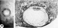
28 A surface view of an intact 12-day ovum after fixation, dehydration and clearing to show the appearance of maternal blood in the lacunar spaces. This is the most distinctive gross feature of and first appears at this stage of development. It is due to flooding of the trophoblastic lacunae with maternal blood. Carnegie 7700, Sequence 9. X 22.
29 A medium-power view of a mid-cross section of the ovum shown in figure 28. Early decidua is developing in the area iimnediately surrounding the ovum. Note three irregular masses of cytotrophoblast—primordial villi—projecting into the syncytium. Note fully developed exocoelomic cavity and membrane with a loose. mesh-work of mesohlasts between it and the cytotroplioblast lining the chorionic cavity. The trophoblast at the abembryonic pole is poorly differentiated, as contrasted to that at the embryonic or implantation pole. Carnegie 7700, Section 6-1-5. X100.
- Streeter Horizon Vc
24-25 surface and mid-cross section 11-day ovum
26-27 surface and mid-cross section 12-day ovum
28-29 surface and mid-cross section 12-day ovum


- Figure Links: 1 | 2 | 3 | 4 | 5 | 6 | 7 | 8 | 9-10 | 11-12 | 13-14 | 15-16 | 17 | 18-19 | 20 | 21-22 | 23 | 24-25 | 26-27 | 28-29 | 30-31 | 32-33 | 34 | 35 | 36 | 37 | 38 | 39 | 40 | 41 | 42 | 43 | 44 | 45 | 46 | 47 | 48 | 40 | 49 | 50 | 51 | 52 | 53 | 54 | 55 | 56 | 57 | 58 | 59 | 60 | 61 | 62 | 63 | 64 | 65 | 66 | 67 | 68 | 69 | 70 | 71 | 72 | 73 | 74 | 75 | 76 | 77 | 78 | 79 | 80 | 81 | 82 | 83 | 84 | 85 | 86 | 87 | 88 | 89 | 90 | plate 1 | plate 2 | plate 3 | plate 4 | plate 5 | plate 6 | plate 7 | plate 8 | plate 9 | plate 10 | plate 11 | plate 12 | plate 13 | plate 14 | plate 15 | plate 16 | plate 17 | table 1 | table 1 image | table 2 image | table 3 image | table 4 | table 4 image | table 5 | table 5 image | All figures | 1956 Hertig | Embryology History - Arthur Hertig | John Rock | Historic Papers
Reference
Hertig AT. Rock J. and Adams EC. A description of 34 human ova within the first 17 days of development. (1956) Amer. J Anat., 98:435-493.
Cite this page: Hill, M.A. (2024, April 27) Embryology Hertig1956 plate04.jpg. Retrieved from https://embryology.med.unsw.edu.au/embryology/index.php/File:Hertig1956_plate04.jpg
- © Dr Mark Hill 2024, UNSW Embryology ISBN: 978 0 7334 2609 4 - UNSW CRICOS Provider Code No. 00098G
File history
Click on a date/time to view the file as it appeared at that time.
| Date/Time | Thumbnail | Dimensions | User | Comment | |
|---|---|---|---|---|---|
| current | 21:27, 23 February 2017 | | 1,280 × 1,904 (452 KB) | Z8600021 (talk | contribs) | |
| 09:58, 23 February 2017 |  | 1,549 × 2,420 (644 KB) | Z8600021 (talk | contribs) | {{Hertig1956 figures}} |
You cannot overwrite this file.
File usage
The following page uses this file: