File:Hertig1956 plate03.jpg

Original file (1,280 × 1,908 pixels, file size: 581 KB, MIME type: image/jpeg)
Plate 3
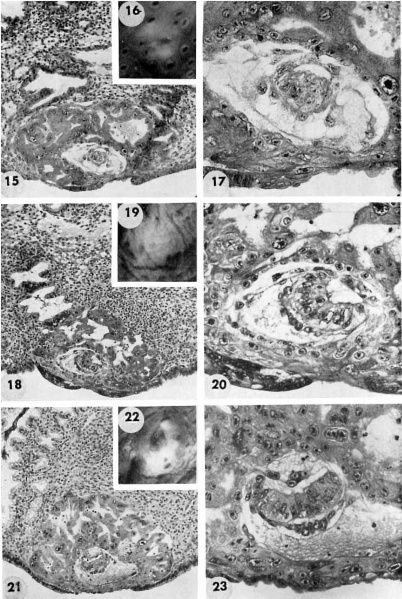
Three 9-day specimens of Horizon Vb, characterized by syncytiotrophoblastic lacunae with early utero-placental circulation, amniogenesis, a simple bilaminar germ disc and only variable degrees of exocoelomie (Heuser’s) membrane formation.
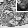
15 Because the decidual reaction is not yet apparent, this ovum is considered to be slightly younger than the other two 9-day specimens illustrated on this plate. The trophoblastic lacunae contain few cellular elements in the plasma. indicating
that at this stage a sluggish circulation begins Within the trophoblastic shell of the ovum. Carnegie 8215, Section 12-5-1-. X100.
16 Surface view of intact specimen. This endomctrium, both grossly and microscopically is slightly younger than that of the following specimens. Carnegie 8215, Sequence 3. X 22.
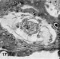
17 Detail of the embryo, chorionie cavity and adjacent trophoblast. Note lensshaped germ disc, amniogenesis from adjacent eytotrophoblast, and delaminating mesoblasts at the abembryonic pole which are continuous with the endoderm and thus together form the exocoelomic membrane. Carnegie 8215, Section 12-54. X 300.
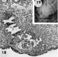
18 Note early predecidual reactions with infiltrating leucocytes about the ovum. Early utero-placental circulation is visible in the form of an empty maternal blood space at right communicating with the coalescing lacunae of the future intervillous space. Carnegie 8171, Section 3-2-11. X 100.
19 The significant feature of this gross implantation site is its inconspicuous nature due to small size of ovum and lack of endomctrial ulceration or congestion. Carnegie 8171, Sequence 3. X 22.
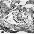
20 Detail of embryo and surrounding trophoblast. Note bilaminar germ disc with vacuolated ectoderm, early amniogenesis and an early exoeoelomic membrane. delaminating from the cytotrophoblast, best seen at the abembryonic pole. Note small shallow surface ulcer with alteration of adjacent nuclei. The paler cytotrophoblast lines the chorionie cavity and varies in thickness whereas the darker synrcytiotrophoblast lies peripherally and contains many intracytoplasmic lacunae which are coalescing to form the future intcrvillous space. Note mitotic figure in cytotrophoblast. Carnegie 8171, Section 3-:2-11. X 300.
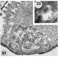
21 The endometrium has slightly more early decidual reaction than 8171 in figure 18. This specimen is similar in general development to 8171 except that its exocoelomic membrane is not yet formed, possibly because of hemorrhage into the chorionic cavity. See figure 23 for details. Carnegie 8004, Section 11-4-7. X 100.
22 Surface view of intact specimen after fixation and partial dehydration. This was prominent prior to fixation because of size, ulceration of endomctrial surface over the ovum and the unusual feature of hemorrhage into the chorionic cavity. This is the youngest human implantation site that was visible without fixation. Carnegie 8004, Sequence 7. X 22.
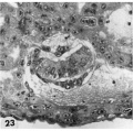
23 Detail of embryo, chorionic cavity and surrounding trophoblast. The maternal hemorrhage in the chorionic and amniotic cavities is abnormal and the result of communication of the trophoblastic lacnmae and chorionic. cavity through a. defective chorionic membrane. A few mesoblnsts are delaminating from the cytotrophoblast but in general this process appears to be delayed perhaps due to the above mentioned hemorrhage. Note mitotic figure in cytotrophoblast. Carnegie 8004, Section 11-4-4. X 300.
- Streeter Horizon Vb
15-16 9-day specimens
17 Embryo, chorionic cavity and adjacent trophoblast
18-19 pre-decidual reaction
20 Embryo and surrounding trophoblast
21-22 endometrium and intact specimen
23 Embryo, chorionic cavity and surrounding trophoblast


- Figure Links: 1 | 2 | 3 | 4 | 5 | 6 | 7 | 8 | 9-10 | 11-12 | 13-14 | 15-16 | 17 | 18-19 | 20 | 21-22 | 23 | 24-25 | 26-27 | 28-29 | 30-31 | 32-33 | 34 | 35 | 36 | 37 | 38 | 39 | 40 | 41 | 42 | 43 | 44 | 45 | 46 | 47 | 48 | 40 | 49 | 50 | 51 | 52 | 53 | 54 | 55 | 56 | 57 | 58 | 59 | 60 | 61 | 62 | 63 | 64 | 65 | 66 | 67 | 68 | 69 | 70 | 71 | 72 | 73 | 74 | 75 | 76 | 77 | 78 | 79 | 80 | 81 | 82 | 83 | 84 | 85 | 86 | 87 | 88 | 89 | 90 | plate 1 | plate 2 | plate 3 | plate 4 | plate 5 | plate 6 | plate 7 | plate 8 | plate 9 | plate 10 | plate 11 | plate 12 | plate 13 | plate 14 | plate 15 | plate 16 | plate 17 | table 1 | table 1 image | table 2 image | table 3 image | table 4 | table 4 image | table 5 | table 5 image | All figures | 1956 Hertig | Embryology History - Arthur Hertig | John Rock | Historic Papers
Reference
Hertig AT. Rock J. and Adams EC. A description of 34 human ova within the first 17 days of development. (1956) Amer. J Anat., 98:435-493.
Cite this page: Hill, M.A. (2024, April 27) Embryology Hertig1956 plate03.jpg. Retrieved from https://embryology.med.unsw.edu.au/embryology/index.php/File:Hertig1956_plate03.jpg
- © Dr Mark Hill 2024, UNSW Embryology ISBN: 978 0 7334 2609 4 - UNSW CRICOS Provider Code No. 00098G
File history
Click on a date/time to view the file as it appeared at that time.
| Date/Time | Thumbnail | Dimensions | User | Comment | |
|---|---|---|---|---|---|
| current | 21:27, 23 February 2017 | | 1,280 × 1,908 (581 KB) | Z8600021 (talk | contribs) | |
| 09:56, 23 February 2017 |  | 1,564 × 2,407 (713 KB) | Z8600021 (talk | contribs) | {{Hertig1956 figures}} |
You cannot overwrite this file.
File usage
The following page uses this file: