File:Hertig1956 plate02.jpg

Original file (1,280 × 2,009 pixels, file size: 482 KB, MIME type: image/jpeg)
Plate 2
Three normal, previllous ova, Streeter Horizon Va, possessing solid syncytio- and cytotrophoblast without lacunar development, an amniotic cavity and/or an early amnion, a simple hilaminar germ disc and early mesoblast formation.
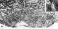
9 A recently implanted ovum of not more than 7.5 days of age, based upon a critical coital date. Note solid trophoblast. composed of paler discrete mononuelear cells and darker less discrete multinucleated cells. The solid disc of trophoblast at the embryonic (implantation) pole is continuous with the membranous unaltered blastocyst wall at the abembryonic pole. Note also the amniogenic cells, tiny cleft-like amniotic cavity, large columnar, non-vacuolated ectodermal cells and smaller tlattcned or polyhedral endodcrmal cells. The ehorionic cavity is irregularly flattened due to collapse of the hlastocyst. Carnegie 8225, Section 20-5-5. X 300.
10 Surface view of the normal 7-day ovum seen in figure 9 photographed under fluid after fixation and partial dehydration. Note embryonic mass within chorionic cavity and the relatively large proportion of the ovum exposed to the uterine lumen. Note prominent gland mouths and surface wrinkling consistent with the 22nd day of the menstrual cycle. Carnegie 8225, Sequence 5. X 22.
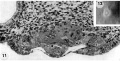
11 Another recently implanted ovum, also not more than 7.5 days of age in view of critical dating of coitus, hut interpreted as being slightly older than 8225 hecause of germ-disc. development. and greater trophoblastic maturity. Note greatest concentration of dark syncytiotrophoblast in the center of the implantation pole dorsal to the embryo. The lighter cytotrophoblast is most prominent peripherally but also lines the inner surface of the proliferating trophoblast. It is amniogenic at its area of contact with the embryo. The nuclei of the surface endomctrial epithelium at its contact with the trophoblast show the abnormality of hydropic degeneration. These might be mistaken for nuclei of syneytiotrophoblastic origin. Actually some of the smaller denser nuclei of the syncytiotrophoblast appear to he of cndometrial stromal origin through the process of ingestion. Carnegie 8020, Section 6-5-9. X 300.
12 Surface view of the same 7-day ovum seen in figure 1. Note clearly visible embryonic mass within dark chorionic cavity which is surrounded by an opaque zone of trophoblast folded at the equatorial plane of the ovum. Carnegie 8020, Sequence 6. X 22.
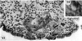
13 An ovum, probably about 8 days of age. Although coital data for this specimen are not critical, its development places it between the 7- and 9-day stages on which critical coital data are available. This inconspicuous stage of implantation is characterized by nearly complete nidation, solid but festooned trophoblast, a small ehorionic. cavity containing a few mesoblasts and a dorsally concave germ disc. Although the primordium of the amniotic cavity is prominent there appears, as yet, to be no amniogencsis. Note actively invading prongs of syncytiotrophoblast surrounding an area of endomctrial stroma and containing two distinct types of nuclei. The large ones divide amitotically but at least some of the smaller ones appear to be of cndometrial stromal origin suggested by the indistinct zone of demarcation between the trophoblast and the maternal stroma. Carnegie 8155, Section 4-4-8. X 300.
14 Surface view of intact ovum shown in figure 13. Note the inconspicuous nature of the slightly raised but opaque implantation site. Carnegie 8155, Sequence 8. X 22.
- Streeter Horizon Va
9-10 implanted ovum 7.5 days of age
11-12 implanted ovum 7.5 days of age
13-14 8 implanted ovum 8 days of age


- Figure Links: 1 | 2 | 3 | 4 | 5 | 6 | 7 | 8 | 9-10 | 11-12 | 13-14 | 15-16 | 17 | 18-19 | 20 | 21-22 | 23 | 24-25 | 26-27 | 28-29 | 30-31 | 32-33 | 34 | 35 | 36 | 37 | 38 | 39 | 40 | 41 | 42 | 43 | 44 | 45 | 46 | 47 | 48 | 40 | 49 | 50 | 51 | 52 | 53 | 54 | 55 | 56 | 57 | 58 | 59 | 60 | 61 | 62 | 63 | 64 | 65 | 66 | 67 | 68 | 69 | 70 | 71 | 72 | 73 | 74 | 75 | 76 | 77 | 78 | 79 | 80 | 81 | 82 | 83 | 84 | 85 | 86 | 87 | 88 | 89 | 90 | plate 1 | plate 2 | plate 3 | plate 4 | plate 5 | plate 6 | plate 7 | plate 8 | plate 9 | plate 10 | plate 11 | plate 12 | plate 13 | plate 14 | plate 15 | plate 16 | plate 17 | table 1 | table 1 image | table 2 image | table 3 image | table 4 | table 4 image | table 5 | table 5 image | All figures | 1956 Hertig | Embryology History - Arthur Hertig | John Rock | Historic Papers
Reference
Hertig AT. Rock J. and Adams EC. A description of 34 human ova within the first 17 days of development. (1956) Amer. J Anat., 98:435-493.
Cite this page: Hill, M.A. (2024, April 27) Embryology Hertig1956 plate02.jpg. Retrieved from https://embryology.med.unsw.edu.au/embryology/index.php/File:Hertig1956_plate02.jpg
- © Dr Mark Hill 2024, UNSW Embryology ISBN: 978 0 7334 2609 4 - UNSW CRICOS Provider Code No. 00098G
File history
Click on a date/time to view the file as it appeared at that time.
| Date/Time | Thumbnail | Dimensions | User | Comment | |
|---|---|---|---|---|---|
| current | 21:26, 23 February 2017 | | 1,280 × 2,009 (482 KB) | Z8600021 (talk | contribs) | |
| 09:53, 23 February 2017 |  | 1,491 × 2,370 (526 KB) | Z8600021 (talk | contribs) | {{Hertig1956 figures}} |
You cannot overwrite this file.
File usage
The following page uses this file: