File:Hertig1956 plate01.jpg

Original file (1,280 × 1,904 pixels, file size: 351 KB, MIME type: image/jpeg)
Plate 1
Normal preimplantation stages in human development; one segmenting egg found in the tube, Streeter Horizon II, and two free intrauterine blastoeysts, Streeter Horizon III.
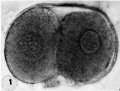
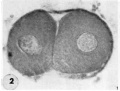
1 Intact two-cell segmenting egg. Note smaller upper polar body between the two blastomeres. 'l‘he lower large one, however, is not in focus. The noun is defective, possibly caused during fixation and partial dehydration or during removal of the ovum from the dish to which it was adherent. See figure 2 for section. Carnegie Embryo 8698, Sequence 12. X 500.
2 Mid-serial section of segmenting tubal egg shown in figure 1. Note nuclear detail, difference in size and shape of blastomeres and loss of zona. Carnegie Embryo 8698, Section 4. X 500.
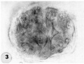
3 An intact 58-cell intrauterine blastocyst. Note eccentrically situated segmentation cavity, the faintly seen inner cell mass and the peripherally arranged blastomeres. The zona and one polar body (lower right) are faintly seen. Carnegie Embryo 8794, Sequence 4. X 500.
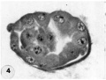
4 Mid-serial section of 58-cell intrauterine blastocyst shown as intact specimen in figure 3. Note absence of zona, about two-thirds of the periphery, probably of artifactitious origin. The two polar bodies are evident at opposite poles of the ovum. The inner cell mass of 5 blastomeres, only 4 of which are present in this section, is lying eccentrically within the segmentation cavity and surrounded by primitive troplioblastic blastomcres. The primitive segmentation cavity, composed of several coalescing spaces, is most clearly delineated as a single unit in this and the two contiguous sections. Note that the nuclei of the embryonic blastomeres, although of equal size, are less vesicular and contain denser clumps of chromatin than those of the trophoblastic blastomeres. Carnegie Embryo 8794, Section 7. X 500.
5 An intact 107-cell intrauterine blastocyst. Note segmentation cavity, cccontrically located inner cell mass at right and pebbly-appearing well formed by trophoblastic blastomerea. There is no zona. Carnegie Embryo 8663, Sequence ‘I. X 500.
6 Mid-serial section of 107-cell blastocyst shown as intact. specimen in figure 5. There are 8 definite large vacuolated formative (embryonic) cells, 69 mural and 30 polar trophoblastic cells. Possibly -1 of the latter are actually formative endodermal cells. Note parts of 4 formative (embryonic) cells at right of picture. The. elongated cell to the upper left of the embryonic disc, interpreted originally as a polar trophoblast, may actually be one of the 4 potential endodermal cells. Note marked irregularity in form and some variation in size of mural trophoblastic cells. The polar trophoblastic cells encircle the. inner cell mass as a corona and vary in shape but tend to be polyhedral. Carnegie Embryo 8663, Section 9. X 500.
7 Endometrium, associated with Carnegie Embryo 8663 (figs. 5, 6), showing the typical morphologic features of the 19th day of a classic 28-day menstrual cycle. Presuma.bly ovulation occurred approximately 108 hours before the surgical removal of the uterus. Note tortuous actively secreting glands, with regularly arranged basal vacuoles and moderately edematous stroma without predecidual reaction. l*‘IlW, S-48-5000. X 91.5.
8 Mucosa of fallopian tube, associated with Carnegie Embryo 8663 (figs. 5. 6). showing the normal tall columnar ciliated epithelium of the mid-interval phase.
1 Two-cell segmenting egg
2 Mid-serial section two-cell
3 Intact 58-cell blastocyst
4 Mid-serial section 58-cell blastocyst
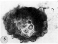
5 An intact 107-cell blastocyst
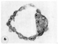
6 Mid-serial section 107-cell blastocyst
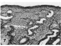
7 Endometrium
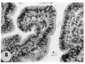
8 Mucosa fallopian tube


- Figure Links: 1 | 2 | 3 | 4 | 5 | 6 | 7 | 8 | 9-10 | 11-12 | 13-14 | 15-16 | 17 | 18-19 | 20 | 21-22 | 23 | 24-25 | 26-27 | 28-29 | 30-31 | 32-33 | 34 | 35 | 36 | 37 | 38 | 39 | 40 | 41 | 42 | 43 | 44 | 45 | 46 | 47 | 48 | 40 | 49 | 50 | 51 | 52 | 53 | 54 | 55 | 56 | 57 | 58 | 59 | 60 | 61 | 62 | 63 | 64 | 65 | 66 | 67 | 68 | 69 | 70 | 71 | 72 | 73 | 74 | 75 | 76 | 77 | 78 | 79 | 80 | 81 | 82 | 83 | 84 | 85 | 86 | 87 | 88 | 89 | 90 | plate 1 | plate 2 | plate 3 | plate 4 | plate 5 | plate 6 | plate 7 | plate 8 | plate 9 | plate 10 | plate 11 | plate 12 | plate 13 | plate 14 | plate 15 | plate 16 | plate 17 | table 1 | table 1 image | table 2 image | table 3 image | table 4 | table 4 image | table 5 | table 5 image | All figures | 1956 Hertig | Embryology History - Arthur Hertig | John Rock | Historic Papers
Reference
Hertig AT. Rock J. and Adams EC. A description of 34 human ova within the first 17 days of development. (1956) Amer. J Anat., 98:435-493.
Cite this page: Hill, M.A. (2024, April 27) Embryology Hertig1956 plate01.jpg. Retrieved from https://embryology.med.unsw.edu.au/embryology/index.php/File:Hertig1956_plate01.jpg
- © Dr Mark Hill 2024, UNSW Embryology ISBN: 978 0 7334 2609 4 - UNSW CRICOS Provider Code No. 00098G
File history
Click on a date/time to view the file as it appeared at that time.
| Date/Time | Thumbnail | Dimensions | User | Comment | |
|---|---|---|---|---|---|
| current | 14:58, 23 February 2017 | | 1,280 × 1,904 (351 KB) | Z8600021 (talk | contribs) | |
| 09:50, 23 February 2017 |  | 1,550 × 2,405 (399 KB) | Z8600021 (talk | contribs) |
You cannot overwrite this file.
File usage
The following page uses this file: