File:Hertig1956 fig83.jpg
Original file (718 × 722 pixels, file size: 68 KB, MIME type: image/jpeg)
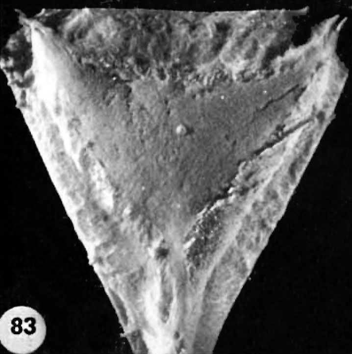
Fig. 83. Abnormal Ova
Two abnormal ova showing malposition of the germ disc and, in one of the specimens, shallow implantation. Whether these ova would ultimately have aborted it is impossible to say.
A surface view of the posterior wall of a uterus showing the position and sim of the implantation site shown in figures 81 and 82. Carnegie 8299
- Figure Links: 1 | 2 | 3 | 4 | 5 | 6 | 7 | 8 | 9-10 | 11-12 | 13-14 | 15-16 | 17 | 18-19 | 20 | 21-22 | 23 | 24-25 | 26-27 | 28-29 | 30-31 | 32-33 | 34 | 35 | 36 | 37 | 38 | 39 | 40 | 41 | 42 | 43 | 44 | 45 | 46 | 47 | 48 | 40 | 49 | 50 | 51 | 52 | 53 | 54 | 55 | 56 | 57 | 58 | 59 | 60 | 61 | 62 | 63 | 64 | 65 | 66 | 67 | 68 | 69 | 70 | 71 | 72 | 73 | 74 | 75 | 76 | 77 | 78 | 79 | 80 | 81 | 82 | 83 | 84 | 85 | 86 | 87 | 88 | 89 | 90 | plate 1 | plate 2 | plate 3 | plate 4 | plate 5 | plate 6 | plate 7 | plate 8 | plate 9 | plate 10 | plate 11 | plate 12 | plate 13 | plate 14 | plate 15 | plate 16 | plate 17 | table 1 | table 1 image | table 2 image | table 3 image | table 4 | table 4 image | table 5 | table 5 image | All figures | 1956 Hertig | Embryology History - Arthur Hertig | John Rock | Historic Papers
Reference
Hertig AT. Rock J. and Adams EC. A description of 34 human ova within the first 17 days of development. (1956) Amer. J Anat., 98:435-493.
Cite this page: Hill, M.A. (2024, April 27) Embryology Hertig1956 fig83.jpg. Retrieved from https://embryology.med.unsw.edu.au/embryology/index.php/File:Hertig1956_fig83.jpg
- © Dr Mark Hill 2024, UNSW Embryology ISBN: 978 0 7334 2609 4 - UNSW CRICOS Provider Code No. 00098G
File history
Click on a date/time to view the file as it appeared at that time.
| Date/Time | Thumbnail | Dimensions | User | Comment | |
|---|---|---|---|---|---|
| current | 14:35, 25 February 2017 | | 718 × 722 (68 KB) | Z8600021 (talk | contribs) |
You cannot overwrite this file.
File usage
The following 2 pages use this file: