File:Hertig1956 fig77.jpg
Original file (1,413 × 997 pixels, file size: 252 KB, MIME type: image/jpeg)
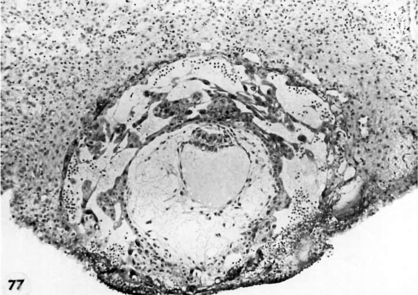
Fig. 77. Abnormal Previllous Ova about 12 to 13 days
A medium-power view of the section through the greatest diameter of an abnormal previllous ovum to show the extreme dilatation of the trophoblastie lacunae. Note the marked hypoplasia of the syncytiotrophoblast which is reduced to fine irregular strands and the relative paucity of cytotrophoblast and the primordial villi resulting therefrom. Note well-formed amnion but relatively small germ disc. The development of the yolk-sac cavity beneath the latter is inconsistent with that of the trophoblast. The primary defect probably lies in the trophoblast with corresponding deficiencies in the structures derived therefrom. Note also the significant deeidual reaction with pronounced leucoeytic infiltration. Inevitable early abortion can be predicted for such a specimen on the basis of its lack of ability to maintain function of the corpus luteum. Such an abortion might take the form of a profuse flow not much later than the next expected period and thus give no undisputed evidence of its true nature. Carnegie 7850, Section 14-3-4. X 100.
- Figure Links: 1 | 2 | 3 | 4 | 5 | 6 | 7 | 8 | 9-10 | 11-12 | 13-14 | 15-16 | 17 | 18-19 | 20 | 21-22 | 23 | 24-25 | 26-27 | 28-29 | 30-31 | 32-33 | 34 | 35 | 36 | 37 | 38 | 39 | 40 | 41 | 42 | 43 | 44 | 45 | 46 | 47 | 48 | 40 | 49 | 50 | 51 | 52 | 53 | 54 | 55 | 56 | 57 | 58 | 59 | 60 | 61 | 62 | 63 | 64 | 65 | 66 | 67 | 68 | 69 | 70 | 71 | 72 | 73 | 74 | 75 | 76 | 77 | 78 | 79 | 80 | 81 | 82 | 83 | 84 | 85 | 86 | 87 | 88 | 89 | 90 | plate 1 | plate 2 | plate 3 | plate 4 | plate 5 | plate 6 | plate 7 | plate 8 | plate 9 | plate 10 | plate 11 | plate 12 | plate 13 | plate 14 | plate 15 | plate 16 | plate 17 | table 1 | table 1 image | table 2 image | table 3 image | table 4 | table 4 image | table 5 | table 5 image | All figures | 1956 Hertig | Embryology History - Arthur Hertig | John Rock | Historic Papers
Reference
Hertig AT. Rock J. and Adams EC. A description of 34 human ova within the first 17 days of development. (1956) Amer. J Anat., 98:435-493.
Cite this page: Hill, M.A. (2024, April 27) Embryology Hertig1956 fig77.jpg. Retrieved from https://embryology.med.unsw.edu.au/embryology/index.php/File:Hertig1956_fig77.jpg
- © Dr Mark Hill 2024, UNSW Embryology ISBN: 978 0 7334 2609 4 - UNSW CRICOS Provider Code No. 00098G
File history
Click on a date/time to view the file as it appeared at that time.
| Date/Time | Thumbnail | Dimensions | User | Comment | |
|---|---|---|---|---|---|
| current | 13:49, 25 February 2017 | | 1,413 × 997 (252 KB) | Z8600021 (talk | contribs) |
You cannot overwrite this file.
File usage
The following 2 pages use this file: