File:Hertig1956 fig55.jpg
Hertig1956_fig55.jpg (635 × 546 pixels, file size: 71 KB, MIME type: image/jpeg)
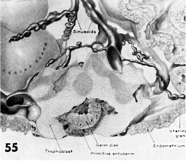
Fig. 55. Reconstruction of half an 8-day ovum
Viewed from the level of the greatest diameter of the embryo. The top section of the reconstruction coincides with the photomicrograph of this ovum in figure 13. The trophoblast (stippled) is still solid and is encircling the tips of maternal siuusoids which, although prominent, actually contain very little blood. The large sinusoid toward the right is interpreted as venous in type, whereas the coiled smaller one to the left is a spiral arteriole. The existing capillary anastomoses between this endometrial arteriole and vcnule are the structures that actually grow and dilate under the stimulus of the immediately adjacent trophoblast. Note amniotic cavity but lack of amnioblasts. dorsally concave hilaminar germ disc and mesoblasts within the chorionic cavity. See figures 13 and 14. Carnegie 8155. X 200.
- Figure Links: 1 | 2 | 3 | 4 | 5 | 6 | 7 | 8 | 9-10 | 11-12 | 13-14 | 15-16 | 17 | 18-19 | 20 | 21-22 | 23 | 24-25 | 26-27 | 28-29 | 30-31 | 32-33 | 34 | 35 | 36 | 37 | 38 | 39 | 40 | 41 | 42 | 43 | 44 | 45 | 46 | 47 | 48 | 40 | 49 | 50 | 51 | 52 | 53 | 54 | 55 | 56 | 57 | 58 | 59 | 60 | 61 | 62 | 63 | 64 | 65 | 66 | 67 | 68 | 69 | 70 | 71 | 72 | 73 | 74 | 75 | 76 | 77 | 78 | 79 | 80 | 81 | 82 | 83 | 84 | 85 | 86 | 87 | 88 | 89 | 90 | plate 1 | plate 2 | plate 3 | plate 4 | plate 5 | plate 6 | plate 7 | plate 8 | plate 9 | plate 10 | plate 11 | plate 12 | plate 13 | plate 14 | plate 15 | plate 16 | plate 17 | table 1 | table 1 image | table 2 image | table 3 image | table 4 | table 4 image | table 5 | table 5 image | All figures | 1956 Hertig | Embryology History - Arthur Hertig | John Rock | Historic Papers
Reference
Hertig AT. Rock J. and Adams EC. A description of 34 human ova within the first 17 days of development. (1956) Amer. J Anat., 98:435-493.
Cite this page: Hill, M.A. (2024, April 27) Embryology Hertig1956 fig55.jpg. Retrieved from https://embryology.med.unsw.edu.au/embryology/index.php/File:Hertig1956_fig55.jpg
- © Dr Mark Hill 2024, UNSW Embryology ISBN: 978 0 7334 2609 4 - UNSW CRICOS Provider Code No. 00098G
File history
Click on a date/time to view the file as it appeared at that time.
| Date/Time | Thumbnail | Dimensions | User | Comment | |
|---|---|---|---|---|---|
| current | 22:43, 23 February 2017 |  | 635 × 546 (71 KB) | Z8600021 (talk | contribs) |
You cannot overwrite this file.
File usage
The following 2 pages use this file:
